
- Title
-
Neurogenin1 is a determinant of zebrafish basal forebrain dopaminergic neurons and is regulated by the conserved zinc finger protein Tof/Fezl
- Authors
- Jeong, J.Y., Einhorn, Z., Mercurio, S., Lee, S., Lau, B., Mione, M., Wilson, S.W., and Guo, S.
- Source
- Full text @ Proc. Natl. Acad. Sci. USA
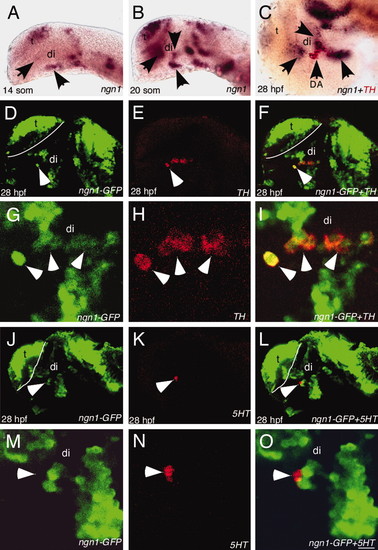
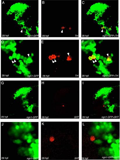
ngn1 is expressed in DA progenitors. All images are lateral views of anterior brain regions. Anterior is to the left, and dorsal is up. (A and B) ngn1 expression at 14- and 20-somite stages, respectively (arrows point to several clusters in the ventral forebrain). (C) A 28-hpf embryo showing ngn1 expression (purple) in close proximity to TH+ DA neurons (red). (D–F) Confocal images of 28 hpf ngn1-GFP transgenic embryos immunostained with GFP antibody (green, D), TH antibody (red, E), and the merged image (F), showing that GFP is detected in TH+ DA neurons. (G–I) High-magnification views of D–F. (J–L) Confocal images of 28-hpf ngn1-GFP transgenic embryos immunostained with GFP antibody (green, J), 5HT antibody (red, K), and the merged image (L), showing that GFP is not detected in 5HT neurons. (M–O) High-magnification views of J–L. di, diencephalon, t, telencephalon. (Scale bars, 64 μm in A and B, 60 μm in C–F and J–L, and 3 μm in G–I and M–O.) EXPRESSION / LABELING:
|
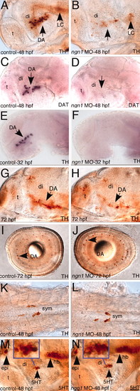
ngn1 is required for DA neuron development in the basal forebrain. All images are lateral views of anterior brain regions except I–L. Anterior is to the left, and dorsal is up. (A–L) Immunostaining with TH antibody (A, B, G, and L), in situ with TH (E and F), or DAT antisense probe (C and D), or immunostaining with 5HT antibody (M and N), shows that DA neurons in the ventral forebrain is completely absent at 32 hpf, largely absent at 48 hpf, and remain significantly defective at 72 hpf in ngn1-morphants, whereas TH+ DA neurons appear normal in the retina, TH+ gut sympathetic neurons are normal in the trunk region of ngn1 morphants, and 5HT neurons are not affected. (Insets) Magnified views of basal forebrain 5HT neurons. DA, dopaminergic neurons; DAT, dopamine transporter; di, diencephalon; epi, epiphysis; hb, hindbrain; LC, locus coeruleus; mhb, midhindbrain boundary; t, telencephalon. (Scale bar, 32 μm.) EXPRESSION / LABELING:
PHENOTYPE:
|
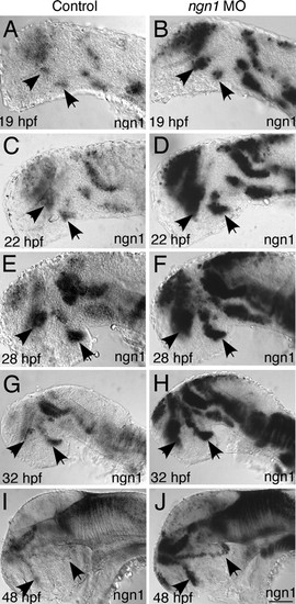
ngn1 expression in ngn1 morphants. Anterior is to the left, and dorsal is up. ngn1 expression is higher in the ngn1 morphants, and Ngn1-expressing progenitor domains are intact in the basal forebrain of ngn1 morphants at various developmental stages as indicated. EXPRESSION / LABELING:
|
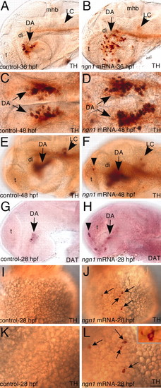
Misexpression of ngn1 leads to supernumerary DA neurons in the forebrain and induces TH+ cells with an apparent neuronal morphology on the yolk surface ectoderm. (A–H) Immunostaining with TH antibody (A–F) and in situ with DAT antisense probe (G and H) shows that DA neurons are significantly increased in the ventral forebrain and ectopically induced in the telencephalon (Di DA neurons are out of focal planes in E–H) in ngn1-mRNA injected embryos. (I and J) Immunostaining with TH antibody showing an ectopic TH+ cell on the yolk surface ectoderm of ngn1-injected embryo. (K and L) A high magnification view of (I and J), and Inset showing an ectopic TH+ cell with an apparent neuronal morphology on the yolk surface ectoderm. DA, dopaminergic neurons; DAT, dopamine transporter; di, diencephalon; LC, locus coeruleus; mhb, midhindbrain boundary; t, telencephalon. (Scale bar, 15 μm in K and L, 32 μm in A–H, and 64 μm in I and J.) |
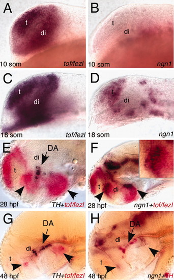
tof/fezl is expressed earlier than ngn1 and later in overlapping domains with ngn1 in the basal forebrain All images are lateral views of anterior brain regions. (A–D) In situ hybridization with tof/fezl cRNA probe (A and C) and ngn1 cRNA probe (B and D). (E–H) Double in situ hybridization of tof/fezl (red)+TH (purple) (E and G), tof/fezl (red) + ngn1 (purple) (F), and TH (red) + ngn1 (pruple) (H). DA, dopaminergic neurons; di, diencephalon; t, telencephalon. (Scale bar, 32 μm in E–H and 64 μm in A and D.) EXPRESSION / LABELING:
|
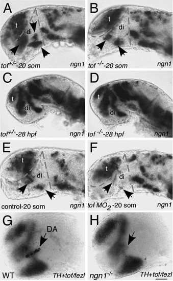
The requirement of tof/fezl in establishing ngn1-expressing DA progenitor domains. (A–D) ngn1 expression in 20-somite wild-type sibling and the tof mutant embryos shows that ngn1 expression in the basal forebrain (arrows) is reduced at 20 somites (A and B) but appears normal at 28 hpf (C and D). (E and F) ngn1 expression is largely absent in the basal forebrain of tof/fezl morphant. (G and H) TH and tof/fezl in situ (both purple) shows that, although TH+ DA neurons are largely absent, the fezl expression appears normal in the ngn1 mutant. di, diencephalon; t, telencephalon. (Scale bars, 32 μm in G and H and 64 μm in A–F.) EXPRESSION / LABELING:
|
ngn1-GFP colocalizes with TH+ dopamine (DA) neurons but not serotonin (5HT) neurons. (A-C) Confocal images of 28 h postfertilization (hpf) ngn1-GFP transgenic embryo immunostained with GFP antibody (green, A), TH antibody (red, B), and the merged image (C). (D-F) High magnification views of A-C. (G-I) Confocal images of 28-hpf ngn1-GFP transgenic embryo immunostained with GFP antibody (green, G), TH antibody (red, H), and the merged image (I). (J-L) High-magnification views of G-I. (Scale bar, 36 mm in A-C, G, and I and 8 mm D-F and J-L.) |