
- Title
-
Retinoids signal directly to zebrafish endoderm to specify insulin-expressing {beta}-cells
- Authors
- Stafford, D., White, R.J., Kinkel, M.D., Linville, A., Schilling, T.F., and Prince, V.E.
- Source
- Full text @ Development
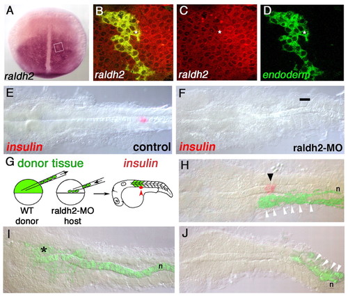
The RA required for pancreas development is produced in anterior paraxial mesoderm. (A) raldh2 expression in dorsal view (anterior to top) at 10 hpf. (B-D) raldh2 expression in endoderm cells; an example is indicated with an asterisk. Confocal images of raldh2 expression (red) at 10 hpf, SOX32-positive donor-derived endoderm is labeled with fluorescein dextran (green). Approximate area of image is boxed in A. (E,F) insulin expression (red) at 24 hpf. (E) Normal insulin expression. (F) No insulin expression appears in raldh2 morpholino injected embryos (bar indicates normal position of pancreas; 100%; n=38). (G) Schematic of cell-transplantation approach. Fluorescein dextran-labeled donor cells from sphere stage (4 hpf) embryos are distributed along the blastoderm margin of raldh2-MO injected hosts. Donor cells contribute to mesoderm (indicated here in anterior somites). (H-J) Transplanted, raldh2-MO injected embryos probed for insulin expression at 24 hpf (red). Photographs are composites of bright field and fluorescent images to detect donor tissue (green). (H) An embryo in which donor tissue contributed to anterior somites (white arrowheads), rescuing insulin expression (arrowhead). (I,J) Embryos in which donor tissue contributed to notochord (n) plus overlying neural cells (*) (I) and posterior somites (white arrowheads) (J); no insulin expression is detectable. |
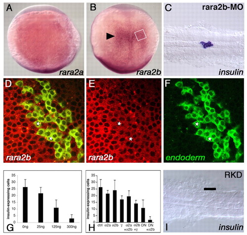
Pancreas development is dependent on RAR function. (A) rara2a expression in dorsal view at 10 hpf. (B) rara2b expression in dorsal view at 10 hpf; arrowhead indicates the approximate AP position where the pancreas will develop. (C) insulin is expressed at 24 hpf following morpholino knock-down of RARα2b (indistinguishable from controls; data not shown). (D-F) rara2b expression in endoderm cells; examples are indicated by asterisks. Confocal images of rara2b expression (red) at 10 hpf, SOX32-positive donor-derived endoderm is labeled with fluorescein (green). Approximate area of image is boxed in B. (G) DN-RARα2a mRNA blocks insulin expression in a dose-dependent fashion (concentrations injected in ng/µl). (H) Histogram showing number of insulin-expressing cells in uninjected control embryos (n=44) compared with embryos injected with RARα2a-MO (n=11), RARα2b-MO (n=12), RARα-MO (n=12), RARα2a-MO plus RARα2b-MO (n=10), RARα2b-MO plus RARα-MO (n=7), DN-RARα2a mRNA (125 ng/ml; n=11) and DN-RARα2a mRNA (125 ng/ml) plus RARα2b-MO (RAR knock-down, RKD; n=85). Error bars indicate standard deviation. (I) Co-injection of DN-RARα2a mRNA and RARα2b-MO blocks insulin expression. |
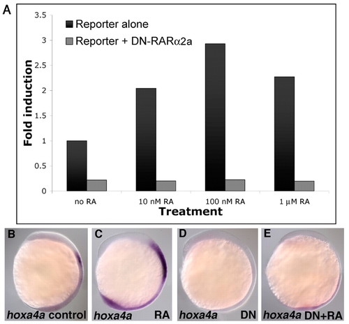
The DN-RARα2a acts in a dominant negative fashion. (A) DN-RARα2a is able to block induction of an RA-responsive luciferase reporter gene (tk-[βRARE]2-luc). Co-injection of DN-RARα2a RNA completely abolishes the response of the reporter gene to RA. Graph shows fold-induction relative to reporter alone; six embryos were used for each experimental condition and assays were carried out in duplicate and averaged. (B-E) hoxa4a expression, 10 hpf, lateral view. (B) Normal embryo. (C) hoxa4a expression is upregulated and extended towards the anterior after 1 hour treatment with RA. (D) DN-RAR mRNA injection blocks hoxa4a expression. (E) DN-RAR blocks the ability of RA to activate hoxa4a expression. EXPRESSION / LABELING:
|
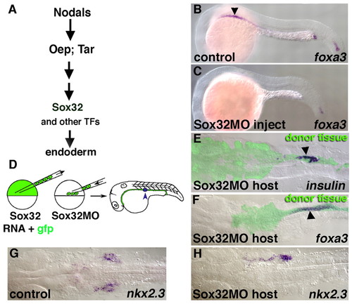
A transplantation technique using SOX32 to target reagents to specific germ layers. (A) Genetic pathway leading to endoderm formation. SOX32 acts downstream of Nodals, their receptors (TAR) and receptor co-factors (OEP) in endodermal specification. (B) Lateral view of foxa3 expression in a 24 hpf control embryo. Endodermal foxa3 expression extends from slightly anterior of the AP level of somite one (arrowhead) towards the posterior, there is also expression in the posterior tip of the notochord. (C) foxa3 is not expressed in the endoderm of SOX32-MO injected embryos, while notochordal expression in the tail is retained. (D) Schematic of the transplantation technique using sox32 to target donor cells to the endoderm. Donor cells from embryos co-injected with SOX32 and GFP mRNA are placed along the blastoderm margin of a SOX32-MO injected host. Following gastrulation, all endoderm is derived from donor cells, which distribute along the ventral AP axis and exhibit regionalized expression of an array of endoderm-specific markers, including insulin (blue arrowhead). (E-H) Transplanted specimens in dorsal view at 24 hpf. (E) insulin expression (arrowhead) in donor-derived endoderm (green). (F) Normal 24 hpf foxa3 expression (arrowhead) in donor-derived endoderm (green). (G) Normal pharyngeal nkx2.3 expression. (H) nkx2.3 expression in donor-derived endoderm; donor tissue has contributed to only the right side in this example but AP patterning is normal. EXPRESSION / LABELING:
|
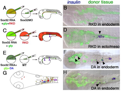
RAR function is required in endoderm but not mesoderm or ectoderm for pancreas development. (A,B) RAR function is required in endoderm for pancreas specification. (A) Schematic illustrating transplantation approach, which is essentially the same as that shown in Fig. 3D, except that donor embryos are additionally injected with RKD reagents at the one-cell stage. (B) Dorsal view of 24 hpf embryo generated as in A; no insulin expression is present (compare with Fig. 3E). (C,D) RAR function is not required in mesoderm or ectoderm for pancreas specification. (C) Schematic illustrating transplantation approach. (D) Dorsal view of 24 hpf embryo generated as in C; insulin expression is present (blue; arrowhead). (E-H) A constitutively active RAR is able to induce ectopic insulin-expressing cells in anterior endoderm. (E) Schematic illustrating transplantation approach; DA is dominant active RAR (xVP16-RARα1). (F,H) Dorsal views of 24 hpf embryos generated as in E. Ectopic donor-derived insulin-expressing cells (blue; arrowheads) are found anterior to the first somite. Photographs are bright-field/fluorescent composite images. (G) Summary diagram showing locations of all insulin-expressing cells in 21 embryos generated as in E; red cells represent embryo in F, blue cells represent embryo in H, yellow cells are from 19 additional specimens. |