
- Title
-
lessen encodes a zebrafish trap100 required for enteric nervous system development
- Authors
- Pietsch, J., Delalande, J.M., Jakaitis, B., Stensby, J.D., Dohle, S., Talbot, W.S., Raible, D.W., and Shepherd, I.T.
- Source
- Full text @ Development
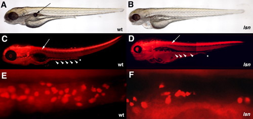
lsn-mutant phenotype. (A,B) Lateral views of live larvae at 96 hpf, showing the abnormal development of the jaw, eye and heart in lsn (B) when compared with wild type (A), and lack of swim bladder in lsn. Arrow in A indicates the swim bladder. (C,D) Lateral views of 96 hpf embryos stained with anti-Hu antibody shows normal DRG development in lsn but a reduced number of enteric neurons that are absent from the distal end of the gut tube. Stars indicate the end of the gut tube; arrows indicate DRGs; arrowheads indicate enteric neurons. (E,F) Lateral views of the gut tube of 96 hpf embryos stained with anti-Hu antibody showing reduced number of neurons in lsn. |
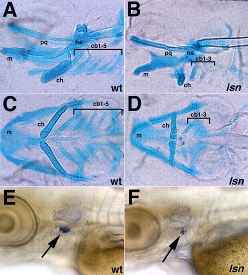
lsn mutation affects both craniofacial and thymus development. (A-D) Alcian Blue staining showing pharyngeal cartilages in 120 hpf wild-type (A,C) and lsn mutant (B,D) larvae shown in lateral (A,B) and ventral (C,D) views. In lsn, the development of the pharyngeal cartilages is abnormal. Notably the ceratobranchials 4 and 5 are absent in lsn. cb 1-5, ceratobranchial cartilages 1-5; ch, ceratohyal cartilage; hs, hyosymplectic cartilage; m, Meckel′s cartilage; pq, palatoquadrate cartilage. (E,F) rag-1 expression at 96 hpf in wild-type (E) and lsn mutant (F) larvae shows that lsn embryos have a significantly reduced thymic primordia (arrows). EXPRESSION / LABELING:
|
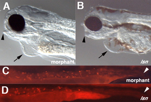
Effect of trap100 antisense morpholino oligonucleotide injection on jaw, heart and enteric neuron development. (A,B) Lateral views of the heads of 96 hpf I>trap100 morphant (A) and lsn mutant (B) embryos. Arrowheads indicate the recessed jaw; arrows indicate cardiac oedema. (C,D) Lateral views of the intestines of 96 hpf embryos stained with anti-Hu antibody. Arrowheads indicate the end of the intestine. The lack of melanophores in morphant the embryo (A) is not due the effect of the trap100 morpholino. Embryos injected with the trap100 morpholino were obtained from an incross of nacre homozygous fish (Lister et al., 1999) and lack melanophores because of a mutation in mitfa. |
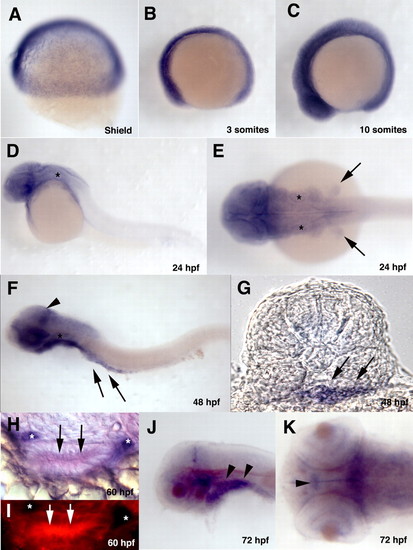
Developmental expression pattern of trap100. (A-F) Whole-mount in situ hybridized embryos hybridized with a trap100 antisense probe at the indicated developmental stages. (A-D,F) Lateral views; (E) Dorsal view of 24 hpf embryo. Asterisks in D-F indicates the otic vesicle. Arrows in E indicate fin buds. Arrowhead in F indicates increased expression of trap100 in the posterior mesencephalon. Arrows in F indicate intestinal mesendodermal expression of trap100. (G) A transverse section taken through 48 hpf embryo at the level of somite 4 showing expression through the intestinal mesendoderm. Arrows indicate intestinal mesendodermal expression of trap100. (H) A transverse section taken trough the gut tube at 60 hpf after double in situ hybridization with a trap100 fluorescein antisense probe (red) and a phox2b digoxigenin antisense probe (purple) showing intestinal epithelia expression of trap100. (I) Same section as in H showing trap100 expression using fluorescence in the intestinal epithelia cells. Arrows and asterisks in H,I indicate intestinal mesendodermal expression of trap100and phox2b-positive ENS precursors, respectively. (J,K) Whole-mount in situ hybridized embryos hybridized with a trap100 antisense probe at 72 hpf. (J) Lateral view; (K) dorsal view. Arrowheads in J indicate pharyngeal arch mesendodermal expression. Arrowhead in K indicates ventral diencephalon cells expressing neurons expressing trap100. In all whole mounts (A-F,J,K), anterior is towards the left; (F,K,J) yolk has been removed. EXPRESSION / LABELING:
|
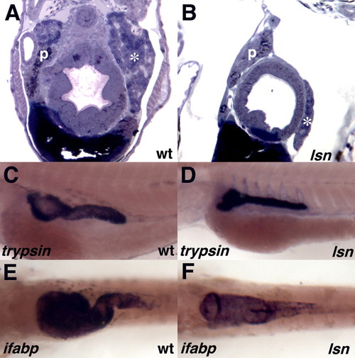
trap100 is required for normal intestinal development but is not required for endoderm-intestine transition. (A,B) Cross-sections of 120 hpf wild-type (A) and lsn mutant (B) embryos stained with Toluidine Blue. (C,D) trypsin expression at 96 hpf in whole-mount in situ hybridized wild-type (C) and lsn mutant (D) embryos. (E,F) ifabp expression at 96 hpf in whole-mount in situ hybridized wild-type (E) and lsn mutant (F) embryos. p, pancreas; *, liver. EXPRESSION / LABELING:
|
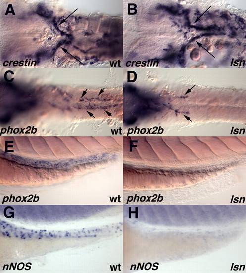
lsn mutation causes a failure of enteric precursors to populate the entire length of the intestine but does not perturb the initial migration of vagal neural crest to the anterior gut. (A,C,E,G) Wild-type and (B,D,F,H) lsn mutants embryos. Anterior is towards the left. (A,B) Ventral view of the vagal region of 36 hpf embryos hybridized with riboprobes for crestin. (C,D) Ventral view of the vagal region to somite 10 of 48 hpf embryos hybridized with riboprobes for phox2b, showing a failure of phox2b-expressing cells to populate the entire length of the intestine. Arrows indicate the migrating enteric precursors. The yolk has been removed from the embryos (A-D). (E-H) Lateral views of the intestine, 72 hpf wild-type (E,G) and lsn mutant (F,H) embryos hybridized with riboprobes for phox2b (E,F) and nos1 (previously nnos) (G,H). EXPRESSION / LABELING:
|
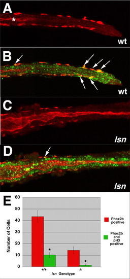
lsn mutants have reduced ENS precursor proliferation. (A-D) Confocal images of dissected intestines from 48 hpf wild-type (A,B) and lsn (C,D) embryos immunocytochemically stained with anti-Phox2b antibody (red) and anti-phosphohistoneH3 antibody (green). (A,C) Anti-Phox2b immunoreactivity in wild type (A) and lsn (C), showing the most posterior point along the gut tube that Phox2b-positive cells can be identified. (B,D) Merged images of the intestines in A and C showing anti-Phox2b and anti-phosphohistone H3 immunoreactivity (arrows). Asterisk in A indicates the end of the gut lumen, which is not opened up to the distal end of the gut tube at this developmental age. Arrows in B,D indicate double-labelled cells. (E) Bar graph showing the mean number of Phox2b immunoreactive cells (red) and double-labelled Phox2b/phosphohistone H3 positive cells (green) present along the entire length of the intestine of 48 hpf wild-type and homozygous mutant lsn embryos. Embryos derived from an incross of heterozygote lsn fish were fixed and stained with anti-Phox2b and anti-phosphohistone H3 antibodies at 96 hpf and genotyped. Numbers represent the mean number of immunopositive cells±s.e.m. for 10 embryos of each genotype. The difference between them was statistically significant (Student's t-test, *P<0.001). EXPRESSION / LABELING:
|
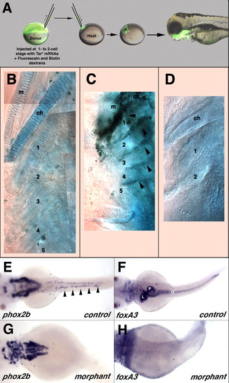
Transplants suggest that lsnw24 functions cell autonomously in the endoderm and endoderm is required for ENS development. (A-D) Transplantation of wild-type endodermal cells partially rescues posterior arch cartilage formation in lsn mutants. (A) At blastula stage, fluorescein-dextran labelled wild-type cells expressing Tar* were transplanted into host and allowed to develop. The lateral view of the head region at 72 hpf shows grafted cells (green) have differentiated into pharyngeal endoderm derivatives. (B-D) Ventral views of 5 dpf larvae stained with Alcian Blue to show cartilages. (B) Wild-type control. All lower jaw cartilages are clearly identifiable. (C) lsn mutant larvae transplanted with wild-type cells showing rescue of posterior ceratobranchial (3-5) in the vicinity of transplanted Tar*-expressing wild type cells (black staining) in the pharyngeal arch endoderm (arrowheads). (D) lsn mutant that lacks posterior ceratobranchials. (E-H) Casanova morphant embryos have no intestinal endoderm and no migrating ENS precursors. Dorsal views of 48 hpf control embryos (E,F) and cas morphant embryos (G,H) hybridized with riboprobes for phox2b (E,G) or foxa3 (F,H) showing a complete lack of phox2b-expressing cells in the region of the intestine of morphant embryos (G) that also completely lack intestinal endoderm (H). Arrowheads in E indicate migrating ENS precursors. m, Meckel's cartilage of the mandibular arch; ch, ceratohyal cartilage; 1-5 ceratobranchial cartilages; G, intestine; L, liver, P, pancreas. EXPRESSION / LABELING:
|
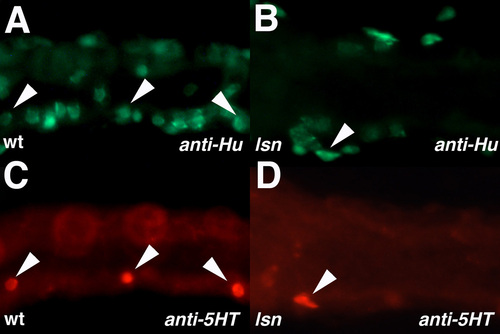
lsn mutants have a reduced number of serotonergic ENS neurons. (A-D) Lateral views of the gut tube of 96 hpf wild type (A,C) and lsn (B,D) embryos that have been double stained with anti-Hu antibody (A,B) and anti-5HT (C,D), showing that there is a reduction in both overall neuron number and serotonergic neurons in lsn mutants when compared with wild type. Arrowheads indicate double-labelled cells. |
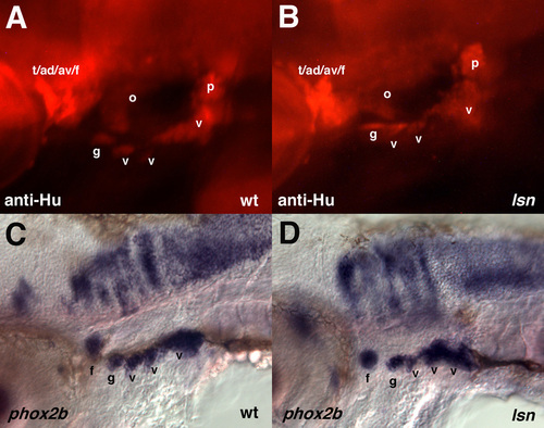
lsn mutants have no cranial ganglia defects.(A, B) Lateral view of Hu expression in cranial ganglia of wild-type (A) and lsn (B) embryos at 96 hpf. (C, D) Lateral flat mounts of embryos showing the cranial ganglia expression phox2b at 48 hpf. ad, anterodorsal lateral line ganglion; av, anteroventral lateral line ganglion; e, enteric neurons; f, facial ganglion; g, glossopharyngeal ganglion; m, middle lateral line ganglion; o, octaval/statoacoustic ganglion; p, posterior lateral line ganglion; v, vagal ganglia. EXPRESSION / LABELING:
|
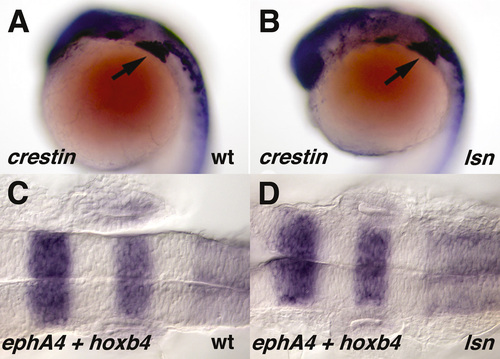
lsn mutants have no defect in initial vagal crest specification and migration or hind-brain patterning. (A, B) Lateral view of crestin expression in wild-type (A) and lsn (B) embryos at 20 hpf. (C, D) Dorsal view of epha4 and hoxb4 expression in wild-type (C) and lsn (D) embryos at 20 hpf. Arrows in A,B indicate vagal crest. EXPRESSION / LABELING:
|
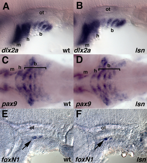
lsn mutants have no defect in the migration and initial differentiation of pharyngeal cartilage neural crest or in initial specification of the thymus.(A, B) Lateral view of dlx2a expression in the migrating neural crest of wild-type (A) and lsn (B) embryos at 36 hpf. (C, D) Ventral views of pax9 expression in the differentiating pharyngeal arch neural crest of wild-type (C) and lsn (D) embryos at 48 hpf. (E, F) Lateral view of foxn1 expression in the thymic primordium of wild-type (E) and lsn (F) embryos at 48 hpf. ot, otic vesicle; b, branchial arches; h, hyoid arch; m, mandibular arch. Arrows in E,F indicate foxn1 expression. |
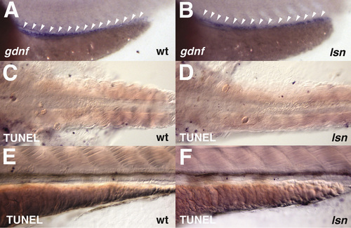
lsn mutants have normal intestinal gdnf expression and have no increase in cell death in the intestine (A, B) Lateral view of gdnf expression in the mesendoderm of wild-type (A) and lsn (B) embryos at 48 hpf. (C, D) Ventral views of the intestines of TUNEL stained wild-type (C) and lsn (D) embryos at 48 hpf. (E, F) Lateral views of the intestines TUNEL stained wild-type (E) and lsn (F) embryos at 72 hpf. EXPRESSION / LABELING:
|

Unillustrated author statements EXPRESSION / LABELING:
|