
- Title
-
Tenascin-C is involved in motor axon outgrowth in the trunk of developing zebrafish
- Authors
- Schweitzer, J., Becker, T., Lefebvre, J., Granato, M., Schachner, M., and Becker, C.G.
- Source
- Full text @ Dev. Dyn.
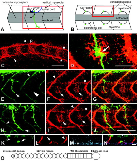
Tenascin-C immunoreactivity is present in the pathway of motor axons in the region of the horizontal myoseptum. A,B: Schematic representations of trunk segments of 22 hours postfertilization (hpf) embryos from a lateral (A) and a dorsal (B) perspective at the level of the horizontal myoseptum give the orientation of C-J and K-N, respectively; rostral is left. Red rectangles indicate the regions shown. rostral (RoP), medial (MiP), and caudal (CaP) primary motor neurons are indicated for one segment in A. C-J: In lateral views of the trunk at 16 hpf (C), tenascin-C immunoreactivity is found in the posterior (p) but not in the anterior (a) half of the somites in addition to vertical myosepta (arrowhead in C) and the ventral and dorsal edge of the notochord (asterisks in C). D-J: At 22 hpf, double labeling of axons (E,H) and tenascin-C (F,I, overlays in G,J) indicates tenascin-C immunoreactivity in the region of the horizontal myoseptum (arrows in F,I) in the caudal, less-mature (E-G) trunk segments, in which axons have not grown out (large arrowhead in E) or have just reached the horizontal myoseptum (arrow in E), and in rostral (H-J) trunk segments, in which axons have passed the horizontal myoseptum (arrow in H). In D, the overlap of tenascin-C (red) and an axon (green) at the horizontal myoseptum (arrow) is shown at high magnification. K-N: In a horizontally rotated image stack, triple labeled for axons (K), tenascin-C (L), and chondroitin sulfates (M, overlay in N), the ventral motor nerve appears as a dot (arrow in K) next to intense tenascin-C immunoreactivity on the lateral surface of a putative sclerotomal cell (arrow in L). Chondroitin sulfates are labeled on the surface of the notochord (asterisk in M) and more weakly on the surface of the sclerotomal cell. C,E,F,H,I,K,L: Vertical myosepta are indicated by arrowheads (C,E,F,H,I,K,L) and notochord by asterisks (C,F,I,M). O: The domain structure of tenascin-C comprises a cysteine-rich region (triangle), 14 epidermal growth factor-like repeats (EGF-like repeats, ovals), 9 fibronectin type III-like domains (FNIII-like domains, rectangles), and a fibrinogen knob (large oval). Scale bar = 50 μm in C, 25 <μm in D, 50 μm in J (applies to E-J), 15.5 μm in N (applies to K-N). EXPRESSION / LABELING:
|
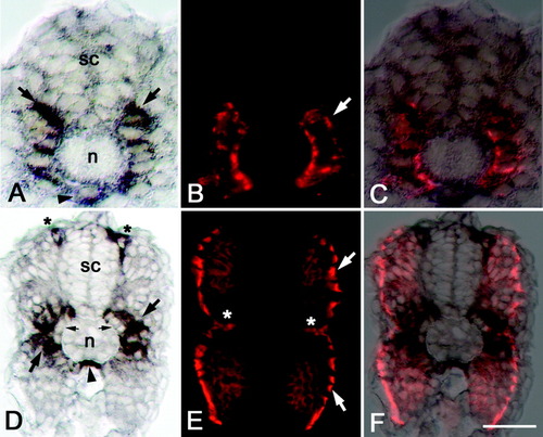
Tenascin-C mRNA is expressed in adaxial cells and other cell types. Cross-sections of trunk regions of zebrafish embryos are shown. Dorsal is up. A-C: At 16 hours postfertilization (hpf), tenascin-C mRNA distribution (A) and immunolabeling for adaxial cell marker F59 (B) overlap (overlay in C) in dorsoventral rows of cells (arrows in A,B) adjacent to the notochord (n) and ventral spinal cord (sc). Hypochord cells (arrowhead in A) also express tenascin-C mRNA. D-F: At 24 hpf, tenascin-C mRNA (arrows in D) is found in weakly F59-immunopositive (E) myotomal cells, putative neural crest cells (asterisks in D), and hypochord (arrowhead in D). Putative migrating sclerotomal cells (small arrows in D) and laterally migrated adaxial cells (arrows in E) do not express the message (see overlay in F), with the possible exception of the muscle pioneer cells (asterisks in E). Scale bar = 25 μm in F (applies to A-F). EXPRESSION / LABELING:
|
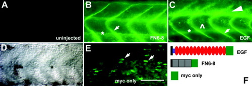
Injections of epidermal growth factor (EGF) repeats RNA and fibronectin (FN) 6-8 RNA lead to secretion of protein. Whole-mounted 24 hours postfertilization (hpf) embryos were labeled with an anti-myc antibody (orientation as in Figure 1A). A,D: Uninjected embryos are unlabeled (A, corresponding differential interference contrast image in D). E: Overexpression of only the myc tag leads to cellular labeling (arrows). B,C: Overexpression of the FN6-8 RNA (B) and the EGF repeats RNA (C) leads to diffuse labeling of extracellular appearance, concentrated in vertical myosepta (arrows in B,C) and around the notochord (asterisks in B,C). C: After injection of EGF repeats RNA, myc immunoreactivity is additionally concentrated in the ventral spinal cord (arrowhead) and in the region of the horizontal myoseptum (open arrowhead). F: Schematic illustrations of RNA constructs: black rectangles, signal peptide; blue rectangle, cysteine-rich region; red ovals, EGF repeats; gray squares, FNIII domains; green squares, myc tag. Scale bar = 50 μm in E (applies to A-E). |
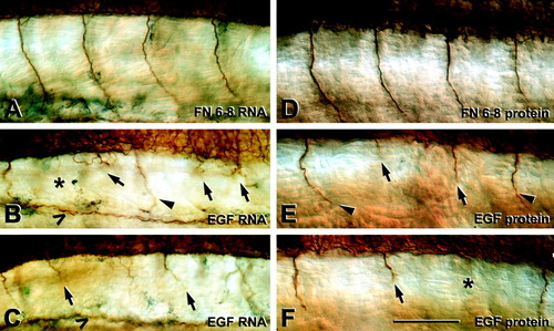
Epidermal growth factor (EGF) repeats RNA and protein injections induce retarded growth of ventral motor nerves. Embryos (24 hpf) were labeled with an anti-tubulin antibody; orientation as in Figure 1A. A,D: After injection of fibronectin (FN) 6-8 RNA (A) or FN6-8 protein (D), ventral motor nerves grow unbranched beyond the horizontal myoseptum. B,C,E,F: Injections of EGF repeats RNA (B,C) or protein (E,F) induce retarded extension of ventral motor nerves (arrows). Asterisks in B,F indicate completely missing nerves. Segments with retarded nerves are interspersed with segments containing apparently normal nerves (arrowheads in B,E). The open arrowheads in B and C indicate additional labeling between the ventral trunk and the yolk extension. Scale bar = 50 μm in F (applies to A-F). |
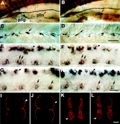
Trunk structures and other axon tracts in 24 hours postfertilization (hpf) embryos are not affected by injection of epidermal growth factor (EGF) repeats RNA. For orientation of A-H, see Figure 1A. In I-L, cross-sections of the trunk are shown (dorsal is up). A-L: Uninjected embryos (A,C,E,G,I,K) were not different from embryos injected with EGF repeats RNA (B,D,F,H,J,L) in tubulin immunohistochemistry (A,B), engrailed immunohistochemistry (C,D), islet-1 (E,F) or islet-2 in situ hybridization (G,H), and F59 (I,J) or F310 (K,L) immunohistochemistry. A,B: The posterior lateral line ganglion (pllg) and nerve (plln) in the anterior trunk region are indicated. C,D: Arrows indicate engrailed immunopositive muscle pioneer cells at the horizontal myoseptum at mid-trunk levels. E-H: In the spinal cord, labeled motor neurons (arrows) and Rohon-Beard cells (arrowheads) are indicated. Islet-1 labels rostral (RoP) and medial (MiP) primary motor neurons. Islet-2 labels caudal primary motor neurons (CaP) and the variably present variable primary motor neurons (VaP). I,J: Arrowheads indicate slow muscle fibers. K,L: Arrowheads indicate fast muscle fibers. Scale bar = 50 μm in D (applies to A-D), 25 μm in H (applies to E-H), 25 μm in L (applies to I-L). EXPRESSION / LABELING:
|
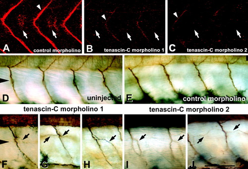
Tenascin-C morpholinos induce aberrant branching of ventral motor nerves. For orientation, see Figure 1A. A-C: At 24 hours postfertilization (hpf), tenascin-C morpholino 1 (B) and tenascin-C morpholino 2 (C), but not control morpholino (A), abolish tenascin-C immunoreactivity in the region of the horizontal myoseptum (arrows) and strongly reduce it in vertical myosepta (arrowhead). D-J: At 33 hpf, aberrant branches of anti-tubulin-labeled ventral motor nerves were observed in embryos injected with tenascin-C morpholino 1 (arrows in F-H) or injected with tenascin-C morpholino 2 (arrows in I,J), but not in embryos injected with control morpholino (E) or in uninjected embryos (D). D,F: Arrowheads indicate the level of the horizontal myoseptum for D,E and F-J, respectively. Scale bar = 50 μm in J (applies to A-J). |
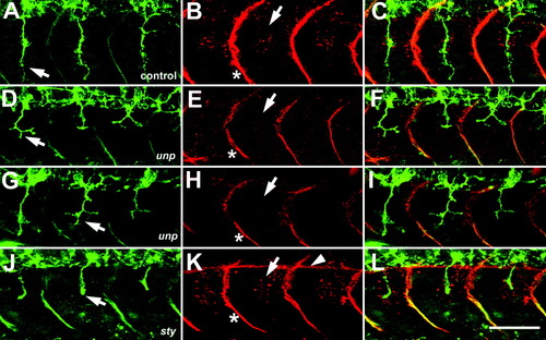
Tenascin-C immunoreactivity is absent from the region of the horizontal myoseptum in unp mutants. A-L: Double immunolabeling of axons (A,D,G,J) and tenascin-C (B,E,H,K, overlays in C,F,I,L) in the trunk at 22 hours postfertilization (hpf); orientation as in Figure 1A. A-C: In a nonhomozygous unp clutch mate, ventral motor nerves are unbranched (arrow in A) and tenascin-C immunoreactivity in the region of the horizontal myoseptum (arrow in B) and in vertical myosepta (asterisk in B) is similar to that in wild-type embryos. D-I: In two unp mutants, motor nerves are stalled and slightly branched (arrows in D,G). E,H: Tenascin-C immunoreactivity is absent from the region of the horizontal myoseptum (arrows) and reduced in vertical myosepta (asterisks) in these embryos. J-L: In a sty mutant, axons stall at the horizontal myoseptum (arrow in J) but tenascin-C immunoreactivity around the horizontal myoseptum (arrow in K) and in vertical myosepta (asterisk in K) is indistinguishable from wild-types. In the image stack in K, the upper surface of the notochord (arrowhead) is included because the embryo is slightly tilted. Scale bar = 50 μm in L (applies to A-L). EXPRESSION / LABELING:
|

Unillustrated author statements EXPRESSION / LABELING:
|