
- Title
-
Molecular cloning and developmental expression of foxP2 in zebrafish
- Authors
- Bonkowsky, J.L., and Chien, C.B.
- Source
- Full text @ Dev. Dyn.
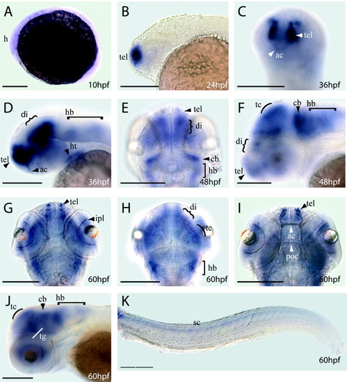
Whole-mount in situ hybridization analysis of foxP2 RNA expression during zebrafish development, stages 10 to 60 hpf. A: Lateral view at 10 hpf. B: Lateral view at 24 hpf. C: Oblique rostral view at 36 hpf. D: Lateral view at 36 hpf. E: Dorsal view at 48 hpf. F: Lateral view at 48 hpf. G,H: Dorsal views at 60 hpf in two different focal planes. I: Dorsal view focused on axon tracts at 60 hpf. J: Lateral view at 60 hpf. K: Lateral view at 60 hpf of expression in the spinal cord. Lateral views: anterior to the left; dorsal views: anterior to the top. Scale bars are 50 μm. ac, anterior commissure; cb, cerebellum; di, diencephalon; h, head region; hb, hindbrain; ht, heart; ipl, inner plexiform layer; poc, post-optic commissure; rgc, retinal ganglion cells; sc, spinal cord; tc, tectum; tg, tegmentum; tel, telencephalon. |
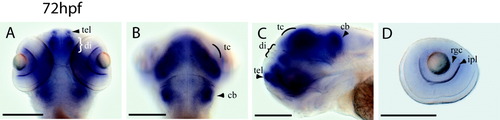
Whole-mount in situ hybridization analysis of foxP2 RNA expression during zebrafish development, stage 72 hpf. A,B: Dorsal views at 72 hpf in two different focal planes. C: Lateral view at 72 hpf. D: Expression in the retina at 72 hpf. Lateral views: anterior to the left; dorsal views: anterior to the top. Scale bars are 50 μm, except in D, where scale bar is 25 μm. cb, cerebellum; di, diencephalon; ipl, inner plexiform layer; rgc, retinal ganglion cells; tc, tectum; tel, telencephalon. EXPRESSION / LABELING:
|
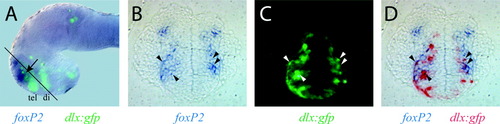
Co-expression of foxP2 and dlx5/6 in the subpallial telencephalon. dlx6a(156i, 156ii):gfp embryos at 32 hpf were double-stained for in situ expression of foxP2 (purple), and with an antibody directed against GFP and detected fluorescently using Alexa-488. A: Whole-mount embryo, lateral view of the head region with dorsal at the top (eyes removed for better visualization). foxP2 expression (purple) overlaps with the telencephalic band of dlx:gfp expression (green, tel) in a narrow area (arrow). Diencephalic expression of dlx:gfp is marked di. Approximate plane of the sections shown in B-D is indicated by the black line transecting the head. B-D: A single 7-μm transverse plastic section in the ventral telencephalon at the level of the rostral olfactory placode, dorsal is at top. Two arrowheads in each hemifield indicate representative cells that show co-expression. B: foxP2 in situ expression. C: GFP expression. D: Overlay of foxP2 and GFP expression, with GFP pattern shown using inverse contrast (in red). EXPRESSION / LABELING:
|