
- Title
-
Cell proliferation in the developing lateral line system of zebrafish embryos
- Authors
- Laguerre, L., Soubiran, F., Ghysen, A., Konig, N., and Dambly-Chaudiere, C.
- Source
- Full text @ Dev. Dyn.
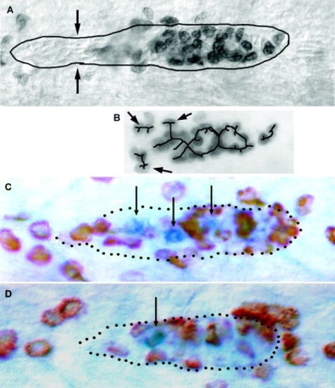
Spatial patterning of proliferation. A: A representative primordium that is four somites away from the last-deposited neuromast. B: The result of the skeletization procedure. Note that the procedure does not take into account the outline of the primordium and considers peridermal cells that abut the primordium (arrows) as linked. This strategy is because we introduced as few constraints as possible in order not to bias the analysis. The pattern defines two full annuli and an incomplete one. Note also the complete depression of replication in the trailing cells, which are about to be deposited (the narrowing marked by the arrows in A indicates that the slowing down of the leftmost cells has already begun). C,D: Two examples of primordia doubly labeled for bromodeoxyuridine incorporation (brown) and for zath1 expression (blue). In both cases, the zath1-expressing cells are surrounded by replicating cells but are themselves nonreplicating. EXPRESSION / LABELING:
|