
- Title
-
Delta-Notch signalling controls commitment to a secretory fate in the zebrafish intestine
- Authors
- Crosnier, C., Vargesson, N., Gschmeissner, S., Ariza-McNaughton, L., Morrison, A., and Lewis, J.
- Source
- Full text @ Development
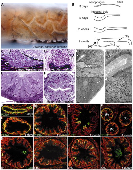
The zebrafish intestine and its pattern of cell renewal. (A) Micrograph of the posterior intestine of a living 2-week-old fish. Intestinal villi are clearly visible through the transparent body wall. The epithelial cells are coloured brown by uptake of pigment in the diet. (B) Sketches showing development of the gross anatomy of the zebrafish gut. A, anterior segment; M, middle segment; P, posterior segment. (C-F) Toluidine Blue-stained plastic sections of intestinal bulb (C,E) and posterior intestine (D,F) at 3 days and 1 month. The intestine matures rapidly from a simple tube to a more elaborate structure with villi. The intestinal bulb initially has a flattened shape (white outline in C). (G-J) Electron micrographs identify goblet cells (G), brush-border cells (H) and enteroendocrine cells (I,J). (J) Enlargement of the area boxed in the top-left corner in I, showing characteristic secretory granules. (K-M) Proliferating cells are progressively restricted to the intervillus pockets as shown by PCNA labelling (green) in the intestinal bulb. (N) Pulse-chase experiment showing the migration of BrdU-labelled cells (green) along the villi of the intestinal bulb 0, 24, 72 and 96 hours after the pulse. (O) TUNEL labelling (green) showing programmed cell death in cells at the tops of the intestinal villi. (P) At 1 month of development, proliferation is more plentiful in the posterior part of the gut, as shown by PCNA labelling (green). (K-P) Confocal optical sections; TOPRO-3 nuclear stain is in red. EXPRESSION / LABELING:
|
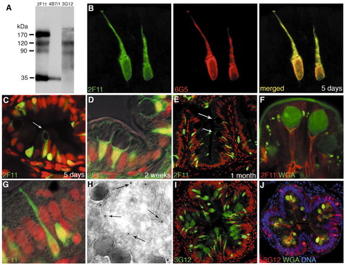
Monoclonal antibodies that mark secretory cells. (A) Western blot of zebrafish gut lysate resolved under non-reducing conditions on SDS-PAGE; antibodies 2F11, 4B7/1 and 3G12 have different antigen specificities. (B) 2F11 (green) and 6G5 (red) stain identical differentiated cells in the intestinal bulb of a 5-day-old larva. (C-H) 2F11 immunolabelling. At 5 days (C), the posterior intestine contains scattered 2F11-positive cells, including a few goblet cells (arrow); goblet cells are more mature and plentiful at 2 weeks (D); by 1 month, goblet cells are also seen in the intestinal bulb (E, arrows). (F) Goblet cells in the intestinal bulb, labelled with wheat-germ agglutinin (green), which stains the mucus compartment, and 2F11 (red), which stains the rest of the cytoplasm. (G) 2F11 (green) also stains some small basal cells and cells that extend thin processes towards the gut lumen; these may be varieties of enteroendocrine cells. (H) Immunogold labelling with 2F11 (small electron-dense particles, arrows) marks the cytoplasm of an enterendocrine cell containing secretory granules (large dark vesicles). (I) 3G12 (green) stains goblet cells in the posterior intestine of a 1-month zebrafish. (J) 3G12 (red) and wheat-germ agglutinin (green) co-stain goblet cells; 3G12 stains both the mucus compartment and the rest of the cytoplasm. Nuclei are stained with TOPRO-3, shown in red (C,D,E,G,I) or blue (J). |
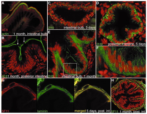
Monoclonal antibodies that mark brush border, epithelial cell polarity and basement membrane in the zebrafish gut. (A,B) 4E8 antibody stains the brush border, identifying absorptive cells. (A) 4E8 (red) recognises an antigen at the apices of the microvilli, distal to the actin-rich microvillus cores (green). (B) The 4E8 stain (green) is specific to absorptive cells; goblet cells (arrows) are unstained. (C-F) 2H9 (green) stains the lateral membranes and basal regions of gut epithelial cells; the staining is already visible at 3 days, and then becomes progressively stronger and more plentiful basally, especially in the villus epithelium, where cells are basally elongated. (F) Detail of the boxed area in E, showing membrane localization; basal stain may be cytoplasmic, or may reflect labelling of highly convoluted plasma membrane. (G,H) 5F11 labels the basement membrane. (G) 5F11 (red) colocalizes with laminin (green) in the intestinal epithelium at 5 days. (H) At 1 month, 5F11 (green) staining outlines the intestinal epithelium. (B-F,H) TOPRO-3 nuclear stain is shown in red. |
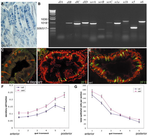
Secretory cells express the Notch ligand Delta and their numbers are increased in aeiAR33, a mutant lacking DeltaD. (A) Intestine of adult mouse heterozygous for a lacZ knock-in at the Delta1 (Dll1) locus. ß-Galactosidase activity (blue) is a reporter for present or past expression of Delta1 expression; the stain is seen in scattered cells, many of which can be clearly identified as goblet cells. (B) Expression of Notch pathway components in zebrafish gut, analysed by RT-PCR. deltaC and deltaD are strongly expressed in the gut, while deltaA and deltaB are not. (C) DeltaD (zdd-2 immunostain in green, arrows) is visible in a subset of secretory cells (2F11, red) in the wild-type zebrafish intestine at 4 days. (D,E) Sections of intestinal bulb of 5-day-old wild-type larvae (D) and aeiAR33 homozygotes (E) stained with 2F11; TOPRO-3 nuclear stain is red. (F) The proportion of 2F11-positive cells is increased all along the length of the gut in aeiAR33 when compared with wild type; data points show mean and s.e.m. from counts of sections of 12 larvae of each genotype. (G) Total numbers of epithelial cells per section for the same set of specimens. |
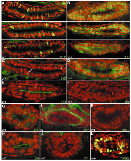
Intestinal phenotype of mib ta52b homozygotes. (A,B) 2F11 staining in the intestinal bulb, comparing three representative pairs of wild-type (A) and mib ta52b (B) specimens at 3 days. The vast majority of epithelial cells are switched to a secretory fate in mib ta52b. (C,D) At the same time, the number of cells expressing DeltaD (zdd-2 antibody, green, indicated by arrows in C) is greatly increased in the mutant. Membrane localization of DeltaD in mib ta52b reflects the failure of Mib-dependent Delta internalization. (E,F) 4E8 staining (green) shows deficit of absorptive cells in three-day mibta52b. (G,H) 2H9 staining (green) shows abnormal epithelial cell polarity in mib ta52b. (I,J) Staining of the basement membrane with 5F11 (green) is also fainter in mib ta52b than in wild-type. (K,L) At 4 days, many dying cells are seen in mib ta52b (L) but not in wild type (K). (A-L) TOPRO-3 nuclear stain is red. EXPRESSION / LABELING:
|