
- Title
-
Supernumerary neuromasts in the posterior lateral line of zebrafish lacking peripheral glia
- Authors
- Lopez-Schier, H., and Hudspeth, A.J.
- Source
- Full text @ Proc. Natl. Acad. Sci. USA
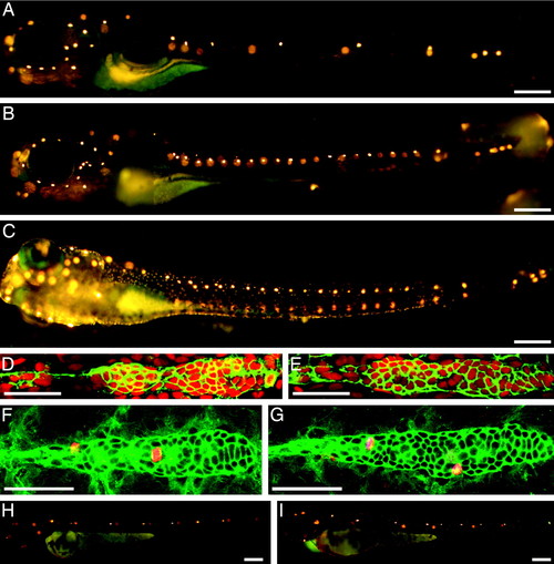
ngn1 mutant zebrafish develop extra neuromasts postembryonically. (A) When treated with the fluorescent compound 4-Di-2-ASP at 6 dpf, a wild-type larva displays only seven to nine neuromasts with functional hair cells on one side of its trunk. The neuromasts on the animal's opposite side appear as diffuse spots of fluorescence. (B) An ngn1 mutant at 6 dpf possesses more than twice as many neuromasts in its posterior lateral line. The number and pattern of neuromasts on the head is essentially normal, as is the animal's pigmentation. (C) At 6 dpf, an animal injected with a morpholino directed against the ngn1 transcript displays a pattern of supernumerary neuromasts comparable to that of an ngn1 mutant. Tilting of the larva separates the images of the neuromasts on the animal's two sides. (D) In a wild-type larva, an antibody against claudin b (green) labels the first primordium and the trail of cells left behind it; the nuclear stain TO-PRO 3 (red) marks all cells. (E) The primordium of an ngn1 mutant does not contain significantly more cells. (F) In a wild-type animal at 24 hpf, antiserum against α-tubulin strongly labels primordial cells (green), whereas that against phosphohistone H3 marks cells in mitosis (red). (G) The primordium is not more active mitotically in a homozygous ngn1 mutant larva. (H) When treated with 4-Di-2-ASP at 48 hpf, a wild-type animal shows only five or six neuromasts with functional hair cells on each side of its trunk. (I) At the same stage, an ngn1 mutant bears an equivalent number of mature neuromasts. In these and all subsequent illustrations, the animal's anterior is oriented to the left and its dorsum is situated to the top. (Scale bars, 50 μm for D–G and 100 µm for A–C, H, and I.) |
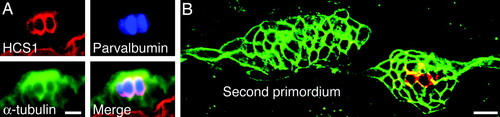
The HCS1 antiserum marks mature hair cells in neuromasts. (A) In a triply labeled wild-type larva, α-tubulin is present throughout the neuromast, whereas parvalbumin 3 and HCS1 occur in mature hair cells. (B) In a wild-type larva at 48 hpf, a neuromast deposited by the first primordium already bears mature hair cells labeled for HCS1 (red). The migrating second primordium is negative for this marker. Both structures are labeled by immunoreactivity against claudin b (green). (Scale bars, 10 μm.) |
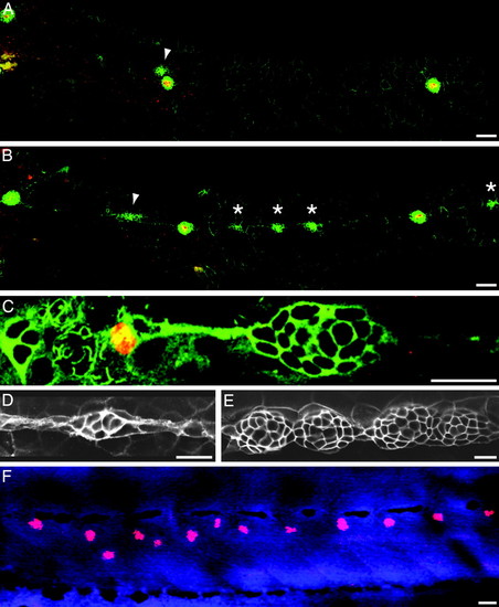
Interneuromast cells form extra neuromasts in ngn1 mutant larvae. (A) In a wild-type larva at 52 hpf, an antiserum against claudin b (green) reveals cells derived from the posterior lateral-line placode. HCS1 labeling (red) marks mature hair cells; the arrowhead designates the migrating second primordium. (B) At the same stage, a homozygous ngn1 larva displays claudin b-positive cell clusters (asterisks) between the mature neuromasts. The arrowhead marks the second primordium. (C) Labeling of an ngn1 mutant embryo at 52 hpf for phosphohistone H3 (red) shows that interneuromast cells remain mitotically active. Labeling for α-tubulin (green) delineates all cells. (D) Labeling for claudin b demonstrates that the interneuromast cells of an ngn1 mutant begin to coalesce at 52 hpf. (E) By 72 hpf, these cell clusters resolve progressively into new neuromasts. (F)An ngn1 mutant larva labeled for HCS1 (red) and TO-PRO 3 (blue) confirms the presence of mature hair cells within the additional neuromasts at 120 hpf. (Scale bars, 10 μm for C–E and 50 μm for A, B, and F.) |
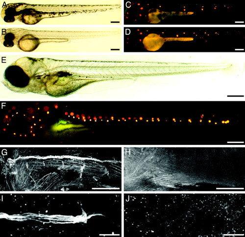
Additional neuromasts also develop in cls mutant larvae. (A) A bright-field micrograph shows a wild-type larva at 48 hpf. (B) A homozygous cls larva displays a similar pace of development. (C) Exposure of the wild-type animal to the fluorescent compound 4-Di-2-ASP reveals the pattern of functional neuromasts. (D) The mutant larva bears an equivalent number of neuromasts at this stage. (E) A bright-field image of a cls mutant larva at 6 dpf demonstrates the absence of pigmentation on the animal′s head and along its body. (F) Treated with 4-Di-2-ASP, the same animal displays 25 neuromasts on the left side of its trunk. However, the number of neuromasts on the head resembles that of a wild-type larva. (G) The marker 6D2 delineates the distribution of glia in the posterior lateral line of a wild-type larva at 72 hpf. (H) At the same stage, a cls mutant lacks peripheral glia along its trunk. (I) 6D2-positive cells occur along the nerve contacting a neuromast in a wild-type larva. (J) No cells of the posterior lateral-line nerve label with the 6D2 antibody in an ngn1 mutant fish. (Scale bars, 100 μm for A–H and 20 μm for I and J.) PHENOTYPE:
|