
- Title
-
tp53 mutant zebrafish develop malignant peripheral nerve sheath tumors
- Authors
- Berghmans,, S., Murphey, R.D., Wienholds, E., Neuberg, D., Kutok, J.L., Fletcher, C.D., Morris, J.P., Liu, T.X., Schulte-Merker, S., Kanki, J.P., Plasterk, R., Zon, L.I., and Look, A.T.
- Source
- Full text @ Proc. Natl. Acad. Sci. USA
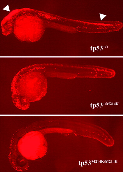
Induction of apoptosis by γ irradiation is suppressed in tp53 mutant embryos. Embryos were γ irradiated at 24 hpf (16 Gray) and fixed at 30 hpf for TUNEL assay. After the assay of multiple embryos resulting from the intercross of tp53M214K mutant heterozygous parents, the embryos were genotyped for their tp53 status. Embryos shown are representative of homozygous wild-type (tp53+/+), heterozygous (tp53+/M214K), and homozygous mutant (tp53M214K/M214K) genotypes. White arrows indicate widespread apoptosis in the brain and spinal cord. Embryos were raised at 28°C. All embryos are shown in lateral view with the head to the left. |
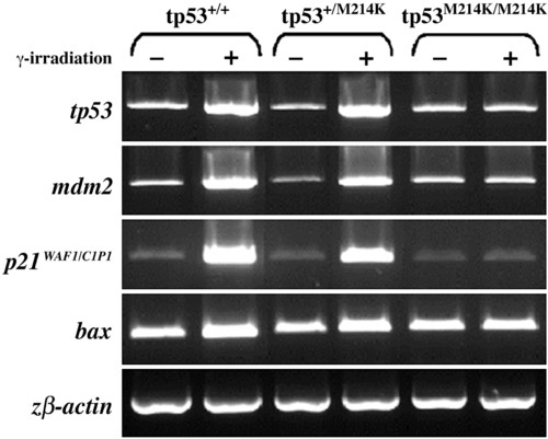
tp53 mutant embryos lack up-regulation of key downstream target genes after γ irradiation. Genes involved in the tp53 regulatory (tp53, mdm2), cell-cycle checkpoint (p21), and apoptotic (bax) pathways were analyzed in homozygous, heterozygous, and wild-type embryos with (+) or without (-) γ irradiation at 24 hpf (16 Gray). Gene expression was assayed at 30 hpf by single-embryo RT-PCR. Results are representative of at least five experiments. β actin was used to control gene expression. EXPRESSION / LABELING:
|
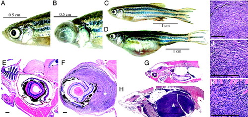
Tumorigenesis features of tp53M214K mutant zebrafish. (A-D) When compared with wild-type zebrafish (A and C), zMPNST development in tp53 mutant zebrafish is identifiable upon external observation because of ocular (B) or abdominal tumor localizations (D). (E-H) When compared with wild-type zebrafish (E and G), histopathology staining with hematoxylin/eosin reveals zMPNST in the eye (F) and abdominal cavity (H), as indicated by the stars (x4). (I-K) Histopathological features of tumors (I) composed predominantly of spindle cells (J) and to a varying degree of epitheloid cells (K) are consistent with the diagnosis of zMPNST. [Bar, 200 μm (E, F, I-K).] |
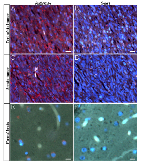
The GFAP expression in primary tumors developed in the zebrafish p53M214K mutant line. Paraffin sections were hybridized with GFAP antisense (A, C, and E) and sense riboprobes (B, D, and F). Expression of GFAP was observed in a periorbital tumor (A, red) and a somite-derived tumor (B, red), compared with the normal levels of GFAP expression in normal brain cortex as a positive control (E, red). Blue indicates nuclear DNA staining with DAPI. (Bar, 40x) EXPRESSION / LABELING:
|