- Title
-
In vivo tracking of T cell development, ablation, and engraftment in transgenic zebrafish
- Authors
- Langenau, D.M., Ferrando, A.A., Traver, D., Kutok, J.L., Hezel, J.P., Kanki, J.P., Zon, L.I., Look, A.T., and Trede, N.S.
- Source
- Full text @ Proc. Natl. Acad. Sci. USA
|
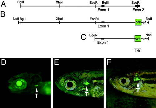
lck-GFP transgenic zebrafish. Diagrams of the genomic DNA sequence comprising the lck promoter (A) and the GFP construct (B) are shown. Enzyme digest sites used for cloning and restriction mapping of the minimal promoter are shown. lck-GFP transgenic fish expressing the 5.5-kb EcoRI-NotI fragment (C) are shown at 8 dpf (D), 45 dpf (E), and 80 dpf (F). Arrowheads denote GFP-labeled cells in the thymus (T). The views are lateral with anterior to the left. EXPRESSION / LABELING:
|
|
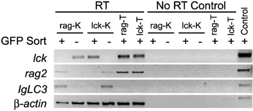
Semiquantitative RT-PCR analysis of FACS-sorted blood cell populations in the kidney (K) and thymus (T) of lck-GFP and rag2-GFP transgenic fish. GFP-positive (+) and -negative (-) blood cell populations are shown. Results for "No RT Control" show absence of genomic DNA contamination in samples. The β-actin PCR control was completed on genomic DNA. Because the β-actin primers span an intron, PCR amplifies a 100-bp larger fragment than seen in RT samples. |
|
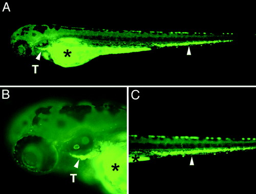
GFP-labeled thymic T cells obtained from adult lck-GFP transgenic fish home to the thymus of transplanted embryos. (A) GFP fluorescent microscopic images of a 4-day-old transplanted embryo (anterior to the left and dorsal to the top) are shown. (B and C) Enlarged views of the head and tail region, respectively. Arrowheads denote GFP-labeled cells in the thymus (T) and in the tail region. Asterisks denote autofluorescence of the yolk sac. |
|
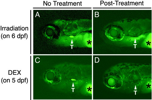
GFP-labeled T cells in 8-day-old lck-GFP fish are ablated in response to γ-irradiation or dexamethasone treatment. (A) Nonirradiated control fish. (B) Fish 2 days postirradiation. (C) Control fish with 0.4% ethanol. (D) Fish 3 days after treatment with 100 μg⋅ml-1 dexamethasone (DEX). Asterisks denote autofluorescence of the yolk sac. Arrowheads denote GFP-labeled cells in the thymus (T). The views are lateral with anterior to the left. |
|
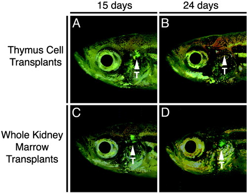
Lymphopoiesis is fully reconstituted in irradiated adult recipients transplanted with lck-GFP kidney marrow. Transplants consisted of thymus cells (1.5 x 10⁶ cells, A and B) or whole kidney marrow (3 x 10⁵ cells, C and D) at 15 or 24 days posttransplantation. The views are lateral with anterior to the left. Arrowheads denote the location of the thymus (T). |
|
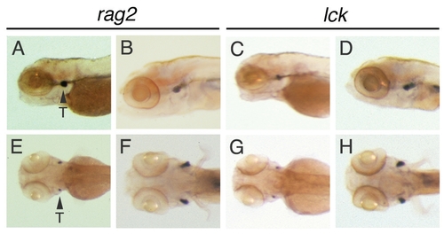
rag2 and lck RNA expression during zebrafish development. In situ hybridization analysis of rag2 RNA expression at 3 dpf (A and E) and 7 dpf (B and F) and of lck RNA expression at 3 dpf (C and G) and 7 dpf (D and H). Lateral views (A–D) and ventral views (E–H) with anterior to the left are shown. Arrowheads denote T cells in the thymus (T, shown only in A and B). |
|
lck RNA expression in zebrafish mutants with hematopoietic defects. Whole-mount in situ hybridization analysis at 4 dpf of wild-type (wt) (A), bloodless (bls) (B), and cloche (clo) (C) fish. Red arrows denote the approximate location of the thymus. Views are lateral with anterior to the left. |
|
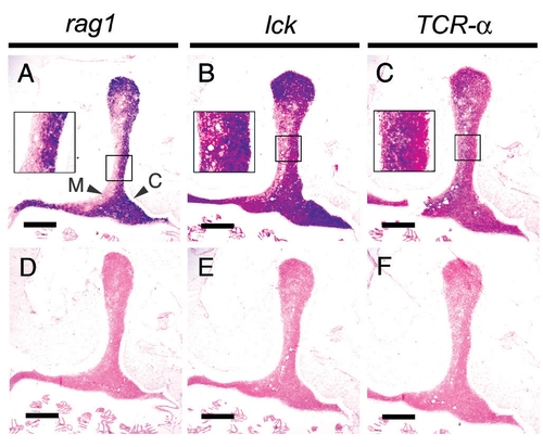
RNA in situ hybridization of paraffin-embedded sections of the thymus. rag1 [antisense probe (A) and sense control (D)], lck (B and E), and TCRα (C and F). Arrowheads denote cortex (C) and medulla (M). (Scale bars, 100 μm.) |
|
Anti-GFP immunostaining of sectioned lck-GFP and rag2-GFP transgenic fish. (A–F) lck-GFP. (G–L) rag2-GFP. Thymus (A and G), kidney (B and H), intestinal lining (C and I), nasal epithelium (D and J), ovary (E and K), spleen (F), and testes (L) are shown. [Scale bars, 100 μm (A and G) and 20 μm (B–F and H–L).] |
|
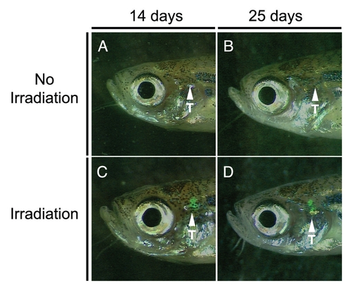
Transplantation of rag2-GFP whole kidney marrow can fully reconstitute the lymphoid system of irradiated recipient fish. Nonirradiated recipient fish transplanted with rag2-GFP whole kidney marrow did not show donor cell engraftment or reconstitution of the lymphoid system as detected by fluorescence microscopy at 14 (A) and 25 (B) days posttransplantation. By contrast, in irradiated recipient fish transplanted with rag2-GFP whole kidney marrow, there is clear engraftment of donor cells and reconstitution of the lymphoid system at 14 (C) and 25 (D) days posttransplantation. Views are lateral with anterior to the left. |

