- Title
-
Ringing in the new ear: resolution of cell interactions in otic development
- Authors
- Riley, B.B. and Phillips, B.T.
- Source
- Full text @ Dev. Biol.
|
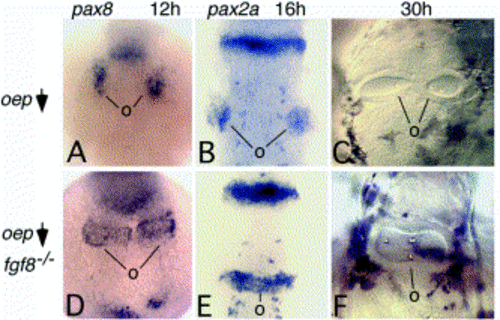
Ear development in embryos deficient in oep and fgf8. (A–C) Wild-type embryos injected with oep-MO as previously described [Phillips et al 2001]. Small bilateral patches of pax8 at 12 h (A) or pax2a at 16 h (B) mark the developing otic placodes (o). By 30 h, otic vesicles form bilaterally but often elongate medially to touch at the midline. However, they are never observed to fuse (over 300 embryos examined). (D–F) ace/+ intercross progeny injected with oep-MO. About 25% of injected progeny showed dramatic changes in gene expression and otic vesicle morphology. These are inferred to be ace/ace(fgf8-/-) mutants. At 12 h, the otic domain of pax8 forms bilateral transverse bands that nearly touch at the midline (D) in 27% (12/44) of embryos. By 16 h, pax2a is expressed in a contiguous transverse stripe through the hindbrain (E) in 19% (9/48) of embryos. At 30 h, a single large otic vesicle forms at the midline and fully spans the width of the hindbrain (F) in 27% (15/55) of embryos. All images are dorsal views with anterior to the top. Abbreviation: o, otic tissue. |
|
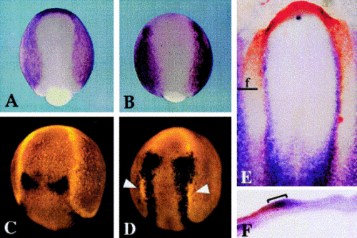
Expression of Dlx and Msx genes in Xenopus, zebrafish, and chick. (A, B) Xenopus embryos at late gastrula stage showing expression of Dlx3 (A) and Msx1 (B). (C, D) Dorsal views of zebrafish embryos at late gastrula or early somitogenesis stages. (C) At bud stage, expression of dlx3 (red) is limited to the preplacodal domain and does not overlap with wnt8 in the hindbrain (black). By the 3-somite stage (D), expression of msxB (black) fully overlaps the dlx3 domain (red) in the preplacodal domain, except in the anterior head and in the lateral portion of the preotic domain (arrowheads). At this stage, msxB expression has started to shift medially into the neural plate. By the 10-somite stage, dlx3 and msxB totally separate into placodal and neural domains, respectively (not shown). (E, F) Chick embryo at stage 6 showing expression of Dlx5 (red) and Msx1 (blue). As seen in a wholemount specimen (E), Msx1 overlaps with Dlx5 except in the anterior head region. The plane of section for (F) is indicated. (F) A section confirms that Dlx5 and Msx1 overlap in the medial preplacodal domain (bracket). Images show dorsal views with anterior to the top (A–E) or a cross section with lateral to the left and dorsal to the top (F). With permission, (A) and (B) are reprinted from Feledy et al. (2001) and (E) and (F) are reprinted from [Streit 2002]. |
|
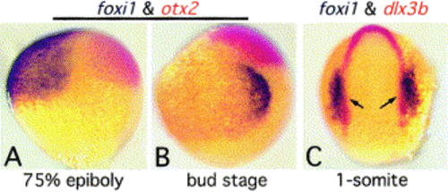
Expression of foxi1 in zebrafish. (A, B) Lateral views (anterior to the top, dorsal to the right) showing expression of foxi1 (black) and the forebrain marker otx2 (red). At 75% epiboly (A), foxi1 is expressed uniformly in the anteroventral quadrant. By bud stage (B), foxi1 has strongly upregulated in preotic placode and downregulated ventrally. (C) Dorsal view (anterior to the top) showing foxi1 (black) and dlx3 (red) at the one somite stage. Expression of foxi1 fully overlaps the preotic domain of dlx3. Reprinted from [Solomon et al 2003] with permission. |
|
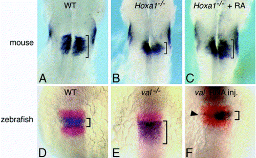
Expression of Fgf3 at comparable stages in mouse and zebrafish. Brackets are shown in all panels to help gauge the size of the Fgf3 domain in the hindbrain. (A–C) Mouse embryos at embryonic day 8.5. (A) A wild-type embryo showing expression in r5 and r6. (B) A Hoxa1-/- mutant shows a greatly reduced hindbrain domain. (C) Brief treatment of Hoxa1-/- mutants with RA restores Fgf3 expression to normal [Pasqualetti et al 2001]. (D–F) Zebrafish embryos at the six-somite stage showing expression of fgf3 (blue) and krox20 (red). (D) Wild-type embryos express fgf3 in r4 and krox20 in r3 and r5. (E) val-/- mutants show loss of krox20 in r5 and expansion of fgf3 into the r5/6 region. (F) A wild-type embryo injected with val mRNA at the 2 cell stage. Expression is primarily restricted to the left side of the embryo where hindbrain expression of fgf3 is nearly extinguished (arrowhead). Expression on the right is essentially normal. Expression of fgf8 in r4 is not altered by either loss of val or misexpression of val [Kwak et al 2002]. With permission, (A–C) are reprinted from [Pasqualetti et al 2001], and (D–F) are reprinted from [Kwak et al 2002]. |
Reprinted from Developmental Biology, 261(2), Riley, B.B. and Phillips, B.T., Ringing in the new ear: resolution of cell interactions in otic development, 289-312, Copyright (2003) with permission from Elsevier. Full text @ Dev. Biol.


