
- Title
-
Cooperative action of ADMP- and BMP-mediated pathways in regulating cell fates in the zebrafish gastrula
- Authors
- Willot, V., Mathieu, J., Lu, Y., Schmid, B., Sidi, S., Yan, Y.-L., Postlethwait, J.H., Mullins, M., Rosa, F., and Peyriéras, N.
- Source
- Full text @ Dev. Biol.
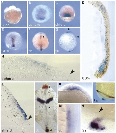
admp expression during early development. In situ hybridization was performed with an admp antisense probe. (A) No signal is detected at the eight-cell stage. (B) Sphere stage, animal pole view. The admp expression domain is radially asymetric. (C) Shield stage, dorsal view, ventral to the top. admp RNA is expressed in the embryonic shield. (D) Sagittal section at 80% epiboly, dorsal to the right, animal pole to the top; admp RNA expression in blue; NTL protein expression in brown. (E) 80% epiboly, dorsal view. admp RNA is expressed along the antero-posterior axis during gastrulation. (F) Tail bud stage, dorsal view. admp expression is discontinuous along the midline. (G) Tail bud stage, lateral view. (H) Sagittal section at the sphere stage, dorsal to the right. An arrowhead indicates the blastoderm margin. (I) Sagittal section through the embryonic shield. An arrowhead shows the blastoderm margin. (J) Flat mount at tail bud stage. Black arrow, hatching gland territory, stained in red with an hgg1 probe; white arrow, midbrain–hindbrain boundary, stained in purple with a pax2.1 probe; black arrowhead, admp RNA staining in the tailbud. (K) Tailbud stage, lateral view; higher magnification of the region marked (+) in (G) and (J). admp RNA is expressed in the prechordal plate. (L) Five-somite stage, lateral view of the tail bud; staining for pax2.1 and admp both in purple. (M) Tailbud stage, lateral view, higher magnification of the region marked (*) in (F, G, J). Axial staining posterior to the midbrain-hindbrain boundary. (N) Five-somite stage; note the Kupffer’s vesicle (arrowhead) next to the admp staining. EXPRESSION / LABELING:
|
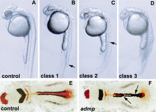
admp overexpression causes the loss of dorsal fates. wt embryos were injected at the one-cell stage with admp RNA (600 pg) and compared to uninjected siblings (control). (A–D) Live embryos at 1 day of development. (E, F) Flat mounts of embryos stained at the one-to three-somite stage with myoD and pax2.1 in dark blue, hgg1, krox20, and ntl in red; anterior to the left. (A) Uninjected embryo. (B–D) admp-injected embryos. (B) Class 1 phenotype. (C) Class 2 phenotype. (D) Class 3 phenotype. (B, C) Arrows indicate the posterior limit of the truncated notochord. (E) Uninjected embryo. (F) admp-injected embryo; notochord defects and fusion of myoD expression domains at the midline are indicated (arrows). |
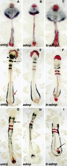
ADMP and TR-ADMP have opposite effects. wt embryos were injected at the one-cell stage with admp RNA (600 pg) or tr-admp RNA (600 pg) and compared to uninjected siblings (uninj.). (A–I) Flat mounts of stained embryos, anterior to the top. (A–C) Flat mounts of embryos stained at 95% epiboly with hgg1, ntl, her5 probes in red and otx2, myoD probes in dark blue. (D–F) Flat mounts of embryos stained at the 10-somite stage with hgg1, ntl, krox20 probes in red and pax6, her5, gata1 probes in dark blue. (D) her5 (black arrow), gata1 (red arrow), and the posterior limit of pax6 expression domain (arrowhead), are indicated. (G–I) Flat mount of embryos stained at the 10-somite stage with ntl, krox20, her5 probes in red and fkd6 probe in dark blue. (G) her5 domain (black arrow) is indicated. (I) End of the notochord is frequently split in tr-admp-injected embryos (arrow). |
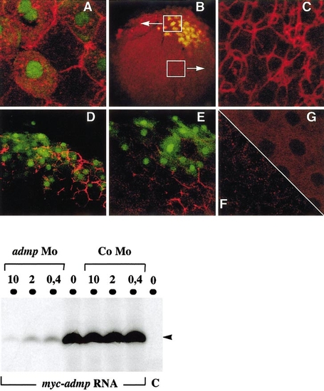
Detection of the MYC-ADMP protein. (Top panel) myc-admp RNA and nls-gfp RNA (50 pg) were injected at the 16-cell stage into 1 marginal blatomere, and MYC-ADMP expression was revealed by immunocytochemistry at the sphere stage. NLS-GFP was used as a lineage tracer to identify the progeny of the injected cell. Embryos where observed with a confocal microscope. (A–C) myc-admp RNA (200 pg) (B)-injected embryo (10x objective); white boxes indicate a GFP-positive region and a GFP-negative region showed at a higher magnification in (A) and (C), respectively. (A, C, G) 63x objective. (A) Injected embryo, GFP-positive domain. (C) Injected embryo, GFP-negative domain. (G) Uninjected embryo. (D–F) 40x objective. (D–E) myc-admp (50 pg) and nls-gfp RNA (50 pg) (E) admp-morpholino (500μM) was injected at the one-cell stage prior to myc-admp and nls-gfp RNA injection at the 16-cell stage. (F) Uninjected embryo. (Bottom panel) myc-admp RNA (1μg) or no RNA (C) was translated in vitro in the absence (0) or in the presence of admp-morpholino (10, 2, or 0.4 μg in a reaction volume of 25 μl). The synthesis of MYC-ADMP protein (45 kDa, arrowhead) was inhibited by the admp-morpholino (admp-Mo) in a dose-dependent manner, and was not affected by the presence of a control morpholino (Co-Mo). |
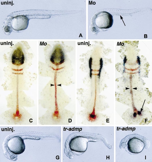
admp-morpholino and tr-admp RNA promote the expansion of dorso-axial structures. wt embryos were injected at the one-cell stage with admp-morpholino (Mo) (250 μM) or tr-admp RNA (600 pg) and compared to uninjected siblings (uninj.). Embryos injected with a control morpholino were unaffected (not shown) and similar to uninjected siblings. (A, B, G–I) Live embryos at 1 day of development. (A) Uninjected embryo. (B) Mo-injected embryos lack the yolk-tube extension (arrow). (C, D) Flat mounts of embryos stained at the three-somite stage with her5, krox20, ntl probes in red and otx2 probe in dark blue. (E, F) Flat mounts of embryos stained at the three-somite stage with her5, krox20, ntl probes in red and fkd6 probe in dark blue. (D, F) Mo-injected embryos have an enlarged notochord (arrowheads). (F) End of the notochord is split in Mo-injected embryos (arrow). (G) Uninjected embryo. (H) tr-admp-injected embryo, A2 phenotype. (I) tr-admp-injected embryo, A3 phenotype. Note the loss of ventral tail fin and vein in tr-admp-injected embryos that is not observed upon Mo injection. PHENOTYPE:
|
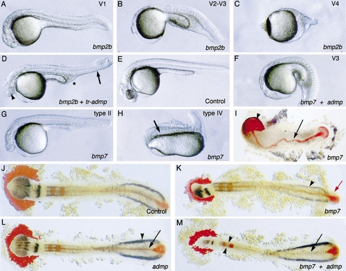
admp, bmp2b, and bmp7 have distinct effects. wt embryos were injected at the one-cell stage with bmp2b, bmp7, admp, or tr-admp RNA either alone or in combination (bmp2b + tr-admp or bmp7 + admp) compared to uninjected siblings (control). (A–H) Live embryos at 1 day of development. (I–M) Flat mounts of embryos stained at the 15-somite stage with pax6, her5, and gata1 in dark blue; hgg1, krox20, and ntl in red; anterior to the left. (A–C) bmp2b RNA-injected embryos with V1, strong V2 (designated V2–V3), or V4 phenotype. (D) Embryos coinjected with bmp2b and tr-admp display both dorsalized (loss of the ventral tail fin and ventral vein indicated by an arrow) and ventralized features (reduced head indicated by an arrow head, excess of blood cells marked “*”). (F) Embryos coinjected with bmp7 and admp display a typical ventralized phenotype. (G, H) bmp7 RNA-injected embryos with type II and type IV phenotype. (H) The notochord is indicated (arrow). (I, K) bmp7-injected embryos. (I) The notochord (arrow) and the hatching gland (arrowhead) are indicated. (K) The prominent tail bud (red arrow) and unaffected gata1 expression domain (arrowhead) are indicated. (L) admp-injected embryo. ntl staining is reduced (arrow) and gata1 is expanded (arrowhead). Compare with (J). (M) bmp7 and tr-admp RNA coinjected embryo. The posterior notochord is lost (arrow) and the neural plate reduced (arrowheads). Numbers and doses corresponding to this experiment are reported in Table 1. |
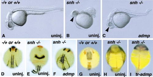
Overexpression of admp or tr-admp RNA and in snailhouse. admp RNA (600 pg) or tr-admp RNA (600 pg) was injected at the one-cell stage into the progeny of snailhouse heterozygous fish. (A–C) Phenotypes were observed live after 1 day of development or (D–I) after in situ hybridization. (D–F) pax 2.1 and myo D in dark blue, hgg1 and krox 20 in red at the five- to seven-somite stage. (G–I) her5 and fkd6 in dark blue, hgg1, krox 20, and ntl in red at the six-somite stage. (A) Uninjected, -/+ or +/+ embryo. (B) Uninjected, snailhouse C4 phenotype. (C) admp-injected snailhouse mutant, C2 phenotype. (B, C) The eye is reduced in (C), compare with (B) (arrowhead). (D, G) Uninjected, -/+ or +/+ embryo. (E) Uninjected, snailhouse C4 phenotype. (F) admp-injected homozygous snailhouse embryo, C2 phenotype. (H) Uninjected, snailhouse C4 phenotype. The neural crest is expanded. (I) tr-admp-injected homozygous snailhouse embryo, C5 phenotype. PHENOTYPE:
|
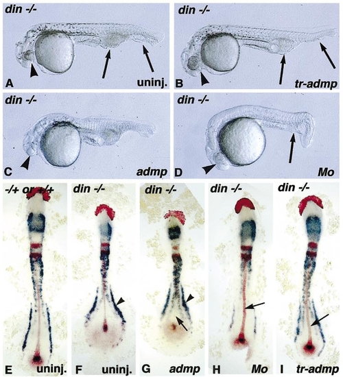
Overexpression of admp, tr-admp, or admp-morpholino in din mutants. tr-admp RNA (600 pg), admp RNA (600 pg), or admp-morpholino (Mo) (250 μM) was injected at the one-cell stage into the progeny of a din-/- x din-/+ cross. (A–D) Phenotypes were observed live after 1 day of development or (E–I) after in situ hybridization at the 10-somite stage; fkd6, her5, and gata1 in dark blue, hgg1, krox20, and ntl in red. (A) Uninjected embryo, din-/- phenotype. (B) tr-admp-injected din-/- embryo, partially rescued. (C) admp injected din-/- embryo, enhanced phenotype. (D) Mo-injected din-/- embryo, partially rescued. (A–D) Arrowhead, compare the size of the eye; arrows, ventral tail fin and vein, lost in (B); and amount of blood cells slightly reduced in (B) and (D). (E) Uninjected, -/+ or +/+ embryo. (F) Uninjected embryo, din-/- phenotype. gata1 expression domain is enlarged (arrowhead) (G) admp injected din-/- embryo, enhanced phenotype. Note the absence of posterior notochord (arrow) and the enlarged gata1 expression domain (arrowhead). (H) Mo-injected din-/- embryo, partially rescued. The notochord is enlarged (arrow); compare with (A) and (B). (I) tr-admp-injected din-/- embryo, partially rescued. The notochord is enlarged (arrow). PHENOTYPE:
|
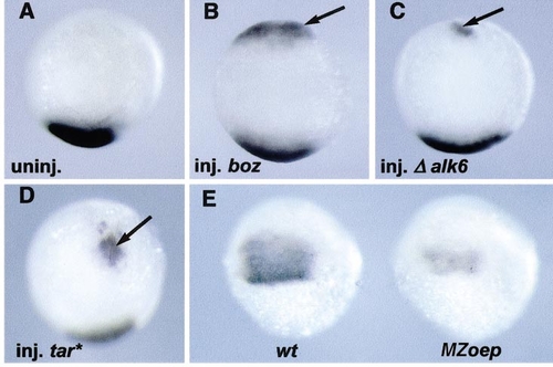
admp induction at the blastoderm ventral margin. (A–E) In situ hybridization with an admp probe at the shield stage. (A–D) Embryos were injected at the 16-cell stage into one marginal blastomere with nls-gfp RNA as a lineage tracer, sorted according to the position of the injection relative to the dorsal side. Ventrally injected embryos were processed for in situ hybridization (animal pole views, ventral to the top). (A) Uninjected embryo. (B) Ventral injection of boz RNA (2 pg). (C) Ventral injection of Δ alk6 RNA (25 pg). (D) Ventral injection of tar* RNA (2 pg). Arrows indicate the induced admp expression domain. (E) Progeny of an oep-/- X oep-/+ cross. Fifty percent of the embryos (MZoep) had a reduced admp expression domain compared with wild-type siblings (wt). EXPRESSION / LABELING:
|
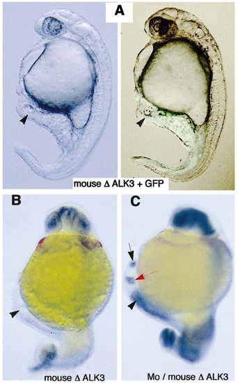
admp antagonizes organizer formation and maintenance. (A, B) Injection of mouse Δ-alk3 RNA at the 16-cell stage into one marginal blastomere leads to induction of a partial secondary axis. Arrowheads, otic vesicles. (A) Bright-field and epifluorescence images are shown side by side. The use of NLS-GFP as a lineage tracer reveals the induced axis. (B, C) In situ hybridization with pax6, her5, and myoD probes in dark blue, hgg1 in red. (C) admp-morpholino injection at the one-cell stage followed by mouse Δ-alk3 RNA injection at the 16-cell stage leads to induction of a partial secondary axis with forebrain structures. The forebrain stained with pax6 (black arrow) and midbrain–hindbrain boundary stained with her5 (red arrow) are indicated. |
Reprinted from Developmental Biology, 241(1), Willot, V., Mathieu, J., Lu, Y., Schmid, B., Sidi, S., Yan, Y.-L., Postlethwait, J.H., Mullins, M., Rosa, F., and Peyriéras, N., Cooperative action of ADMP- and BMP-mediated pathways in regulating cell fates in the zebrafish gastrula, 59-78, Copyright (2002) with permission from Elsevier. Full text @ Dev. Biol.