
- Title
-
Zebrafish early macrophages colonize cephalic mesenchyme and developing brain, retina, and epidermis through a M-CSF receptor-dependent invasive process
- Authors
- Herbomel, P., Thisse, B., and Thisse, C.
- Source
- Full text @ Dev. Biol.
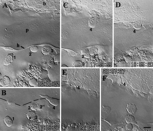
Early macrophage settling in a branchial arch in the 26- to 30-somite period (22–24 hpf). Sequential lateral views of the same embryo, dorsal upward, rostral to the left. (A) Numbers mark three young macrophages at the lateral border of the pericardial envelope (p), outlined by the pericardial hinge (h, see Fig. 3A in Herbomel et al. 1999). (B) Cell 2 divided; a new young macrophage (4) is climbing on the dorsal side of the pericardium. (C) It now reaches the edge of the branchial arch, (D) progressively enters it, (E) and settles there. (F) Fifteen minutes later, two other young macrophages climbed on the dorsal pericardium by the very same route, but then stayed there, becoming residents of the dorsal pericardium. Time indicated in h, min, s; (b) branchial arches, (y) yolk. Bar, 10 μm. |
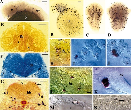
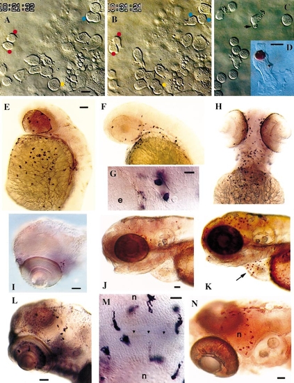
Rapid spreading of early macrophages throughout the head. (A–G) In situ hybridization for L-plastin. (A) Lateral view of the head, 25 hpf. (B) Lateral view of the head and yolk sac, 35 hpf. (C, D) Dorsal views of the head at 35 hpf (C) and 48 hpf (D). (E–G) Cross sections; (E) 25 hpf; two macrophages in the interstices between eyes and forebrain (fb); (F) 35 hpf; three macrophages adhering to the ventral basal lamina of the cerebellum anlage (cb); (G) 35 hpf; macrophages caudal to the otic vesicle, migrating on the hindbrain walls (arrows) and inside the hindbrain ventrally. (H–N) Live embryos at 28–35 hpf viewed by DIC combined with neutral red staining (H–M), or by DIC alone (N); lateral views, dorsal up, anterior to the left. (H–J) Macrophages in the yolk sac blood circulation valley, showing the progression of the neutral red staining process (see text); cells with no stained lysosomes are erythroblasts. (K) Two probably sister macrophage cells adjacent to the otic vesicle. (L) Three macrophages associated with the trigeminal ganglion (tg). (M) Macrophage wandering on the roof of the fourth brain ventricle (v). (N) Phagocytic macrophage (arrow) among trigeminal ganglion neurons. (y) yolk sac; (e) eye; (ov) otic vesicle; (fb) forebrain; (hb) hindbrain; (cb) cerebellum anlage; (tg) trigeminal ganglion; (v) fourth brain ventricle. Bars: (A) 50 μm; (B–D) 50 μm; (E) 50 μm; (F, G) 25 μm; (H–K) 10 μm; (L–N) 10 μm. |
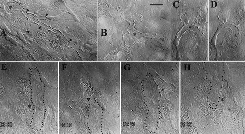
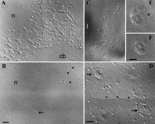
Early macrophages in the yolk sac epidermis. Asterisks mark macrophage nuclei. Filaments inside cells are mitochondria. (A, B) 30 hpf; macrophages of the still type, interdigitating (black arrowheads in A) between epidermal cells. (C, D) 30 hpf; macrophage wandering round a small epidermal cell; (D) is 80 s after (C). (E–H) 48 hpf; macrophage wandering all the way round a long epidermal cell (dotted contour) in 12 min. Time indicated in min, s. Bar, 10 μm. |
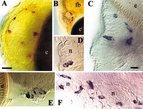
Early macrophages around olfactory organs and neuromasts. (A, B) Neutral red vital staining of macrophages associated with the left olfactory organ at 50 (A) and 84 hpf (B, low magnification). (C–F) In situ hybridization for L-plastin; (C) Right olfactory organ, 96 hpf, dorsal view. (D) Two macrophages, probably sister cells from a recent mitosis, adjacent to an anterior neuromast at 120 hpf. (E) A latero-ventral neuromast just caudal to the eye at 120 hpf. (F) Two neuromasts of the supra-orbital line at 144 hpf. (fb) forebrain; (e) eye; (n) neuromast. Bars: (A) 20 μm; (C–F) 10 μm. |
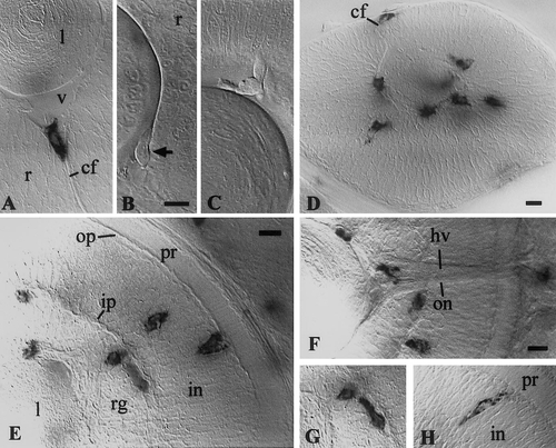
Early macrophages in the retina. (A, D–H) In situ hybridization for L-plastin. (B, C) Live embryos, 30 hpf. (A, B) macrophage in the choroid fissure, entering the vitreous space, between lens and retina, at 26 (A) and 30 hpf (B). (C) macrophage already in the vitreous space. (D) 35 hpf; macrophages have invaded the vitreous-proximal part of the retina; one is appearing in the choroid fissure. (E–G) 48 hpf; the macrophage lying tangentially along the inner plexiform layer in (E) is shown in focus in (G); (H) 72 hpf. (v) vitreous space; (l) lens; (r) retina; (pr):photoreceptor cell layer; (op) outer plexiform layer; (in) inner nuclear layer; (ip) inner plexiform layer; (rg) retinal ganglion cell layer; (cf) choroid fissure; (on) optic nerve; (hv) hyaloid vein. Bars, 10 μm. |
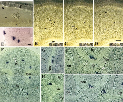
Early macrophages in the brain. (A–D) Live embryos, 35 hpf, lateral view, rostral to the left. (A) Two macrophages in the fourth brain ventricle, dorsal to the first somite. (B–D) Three instants of the macrophage wandering in hindbrain rhombomere 5 (shown in movie 2; asterisk, macrophage nucleus; two neuroepithelial cell nuclei labeled by black dots serve as reference points; time indicated in h, min,s. (E–J) In situ hybridization for L-plastin. (E) 35 hpf, hindbrain, left half, dorsal view, rostral upward. (F–J) Resin cross sections, 48 hpf. (F) Section through the pretectum (pt), hypothalamus, and pituitary (p). (G) Macrophage in the tract of the post-optic commissure (TPOC). (H) Macrophage partly in the tegmentum (tg), partly in the sub-tegmental commissure (stc). (I, J) Two successive sections, in rostro-caudal order, showing one macrophage bridging the optic chiasm (oc) and post-optic commissure (poc) (I, J), and another one in the mesenchyme, riding on the optic nerve (J). (h) hindbrain wall; (v) fourth ventricle; (rf) roof of the fourth ventricle; (ep) epidermis; (ov) otic vesicle, (hy) hypothalamus, (tpoc) TPOC, (r) retina, (on) optic nerves. Bars, 10 μm. |
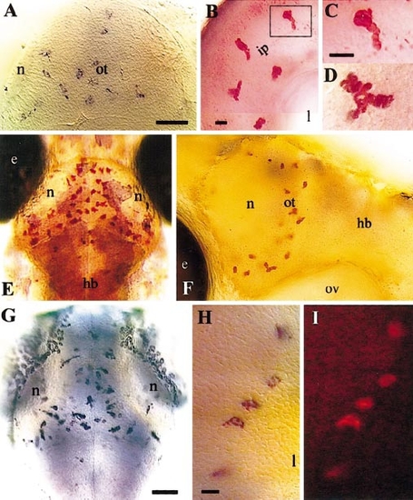
Phenotypic transition of brain and retinal macrophages to early microglia; colonization of the optic tectum. (A, E, G) Dorsal view of the optic tectum, rostral upward. (A) 60 hpf, L-plastin in situ hybridization. (B–F) Neutral red-stained live embryos. (B) Retina, 65 hpf; the boxed early microglial cell is shown at higher magnification in (C). (D) Optic tectum, 82 hpf. (E, F) 99 hpf; (F) lateral view showing the extent of the left tectal neuropile, (G) In situ hybridization for apoE, 72 hpf. (H, I) Retina, 72 hpf; double in situ hybridization for L-plastin (blue) and apoE (red), (I) showing the red fluorescence of the apoE signal. (hb) hindbrain; (ot) optic tectum; (n) tectal neuropile; (e) eye; (ip) inner plexiform layer; (l) lens; (ov) otic vesicle. Bars: (A) 50 μm; (B) 10 μm; (C, D) 10 μm; (G) 50 μm; (H, I) 10 μm. |
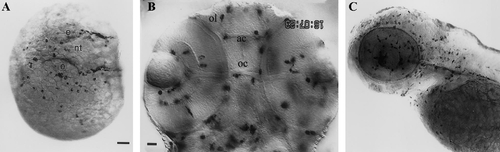
Expression of the fms gene in the early macrophage lineage. (A) 25-somite (21.5 hpf), dorso-lateral view; beside pre-/young macrophages spread on the yolkball, fms-positive xanthophore precursors outline the lateral borders of the neural tube (nt); (e) eye. (B) 48 hpf, head, deep dorsal view. Out-of-focus fms-positive macrophages produce the multiple shadows. (ol) olfactory organ; (ac) anterior commissure; (oc) optic chiasm. (C) 72 hpf, lateral view. Bars: (A, C) 50 μm; (B) 20 μm. |
Behavior of early macrophages in the panther mutant. All panther embryos, except K. (A–D) Live recordings of early macrophage behaviors in the yolk sac. (A, B) 26-somite stage (22 hpf). (A) Red dots mark sister cells from a mitosis that occured 15 min earlier; two other cells are presently in mitosis: the blue-dotted cell in anaphase B, the yellow-dotted one in pro-metaphase. (B) Ten minutes later, the blue-dotted cell cleaved in two daughter cells; the yellow-dotted cell is in anaphase. (C) 25 hpf; erythroblasts (rounded cells) have now joined the yolk sac and macrophages interact with them; one is phagocytosing an apoptotic residue (arrow). (D) 25 hpf, neutral red staining. (E–H) In situ hybridization for L-plastin. (E, F) Lateral views at 36 hpf (E; compare with wt in Fig. 2B) and 44 hpf (F). (G) 44 hpf, dorsal view, anterior to the left; two positive cells in a cephalic vessel. (H) 72 hpf, ventral view. (I, L) In situ hybridization for apoE at 72 hpf (I; compare with wild-type in Fig.7G) and 132 hpf (L); (J, K) 72 hpf; neutral red staining of live panther (J) and wild-type (K) embryos, showing macrophages associated with the optic tectum and regressing hatching gland (arrow). (M) 132 hpf; in situ hybridization for apoE, optic tectum, dorsal view, rostral to the left; (arrowheads) midline. (N) 7.5 days, neutral red staining (compare with wild-type at 99 hpf in Fig. 7F). (n) tectal neuropile. Bars: (A–C) 10 μm; (D) 5 μm; (E, F, H) 50 μm; (G) 10 μm; (I) 50 μm; (J, K) 50 μm; (L) 50 μm; (M) 20 μm; (N) 50 μm. |
Apoptotic bodies accumulate in panther optic tectum and retina until eventual microglial colonization. Live embryos, 120 hpf, DIC optics. (A, B) Optic tectum, dorsal view, rostral upward, (A) Panther embryo. (B) Wild-type embryo; yellow shadows are due to xanthophores present above the brain (absent in panther); (arrow) blood vessel; (arrowheads) mitotic cells. (C) Retina. (D) Newly arrived early microglial cells (arrows) eliminating apoptotic bodies in the optic tectum; (arrowheads) midline. (E, F) Close-up on a tectal phagocyte at two depth planes, showing 7 still recognizable apoptotic bodies within its phagocytosed material; (asterisk) macrophage nucleus. (cb) cerebellum, (n) tectal neuropile. Bars: (A–C) 10 μm; (D) 10 μm; (E, F) 5 μm. |
Reprinted from Developmental Biology, 238(2), Herbomel, P., Thisse, B., and Thisse, C., Zebrafish early macrophages colonize cephalic mesenchyme and developing brain, retina, and epidermis through a M-CSF receptor-dependent invasive process, 274-288, Copyright (2001) with permission from Elsevier. Full text @ Dev. Biol.