
- Title
-
Too much interference: injection of double-stranded RNA has nonspecific effects in the zebrafish embryo
- Authors
- Oates, A.C., Bruce, A.E., and Ho, R.K.
- Source
- Full text @ Dev. Biol.

tbx16/spadetail dsRNA injection into zebrafish embryos appears to cause a mosaic phenocopy of the spadetail mutation. Embryos in (b–d, h) are viewed from the animal pole, and those shown in (f, g) are viewed from the dorsal side. (a) Regions of the spadetail mRNA used to produce nonoverlapping antisense riboprobe and ss or dsRNA transcripts. (b–d) Expression of endogenous spt mRNA in response to injection of tbx16/spt dsRNA. Embryos are shown in animal pole view at the 40% epiboly stage after in situ hybridization with a riboprobe specific for the portion of the tbx16/spt transcript encoding the T-box. (b) Embryos injected with 40 pg tbx16/spt dsRNA; sectors devoid of endogenous spt mRNA are highlighted with arrowheads. (c) Expression of endogenous spt in the wild-type embryo (wt) and widespread loss in the spt mutant embryo (spt) at 40% epiboly. (d) Endogenous tbx16/spt expression at 40% epiboly is unaffected by injection of antisense tbx16/spt ssRNA corresponding to the same region of the mRNA used to generate dsRNA. (e) Dose response to tbx16/spt dsRNA injection measured by depletion of endogenous tbx16/spt mRNA at 40% epiboly. Different concentrations of RNA were injected into embryos at a constant volume. Between 20 and 56 embryos were used for each concentration point. (f) Formation of anterior trunk somites in response to tbx16/spt dsRNA. Embryos at the 3-somite stage, showing normal somite boundary formation, denoted with arrowheads in wild-type, uninjected embryos (wt), unilateral loss of anterior somites in tbx16/spt dsRNA-injected embryos (dsRNA), and complete lack of somites in spt mutant embryos (spt). (g) Expression of red blood cell marker gata1 in 10-somite embryos, showing normal primitive blood in wild-type, uninjected embryos and a depletion of gata1-expressing cells along the lateral plate mesoderm (arrowheads) in tbx16/spt dsRNA-injected embryos. (h) Expression of prospective paraxial mesoderm marker papc at 50% epiboly with dorsal to the top, showing radial expression in the margin in wild-type, uninjected embryos, disrupted marginal expression in tbx16/spt dsRNA-injected embryos, and an absence of papc in the margin in spt mutant embryos. |
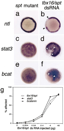
The effects of tbx16/spadetail dsRNA injections are not restricted to those seen in the spadetail mutant. Embryos are viewed from the animal pole at 40% epiboly after in situ hybridization with ntl (a, b), stat3 (c, d), or β catenin riboprobe (e, f). Embryos from spt mutant clutches exhibit gene expression patterns identical to those of wild type (a, c, e), whereas injection with 40 pg tbx16/spt dsRNA causes depletion of the message from sector-like regions of the blastoderm (b, d, f). (g) Concentration dependence of the depletion of endogenous tbx16/spt, stat3, and β catenin mRNA at 40% epiboly on the amount of injected tbx16/spt dsRNA, measured by in situ hybridization. |
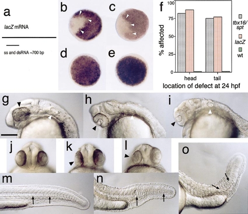
dsRNA causes nonspecific depletion of mRNA. Embryos are viewed from the animal pole in (b–e), from a lateral perspective with anterior to the left in (g–i) and (m–o), and from a rostral perspective in (j–l). Scale bar for (g–o) is 250 μm. (a) Region of the E. coli lacZ mRNA used to produce sense, antisense, and dsRNA preparations. (b–e) Expression patterns of endogenous tbx16/spt and β catenin in embryos at 40% epiboly after injection of either ss or dsRNA from the lacZ gene. spt (b) and β catenin (c) expression after lacZ dsRNA injection was depleted from sector-like regions of the blastoderm in a manner identical to that seen after 40 pg tbx16/spt dsRNA injection. Injection of sense (d) or antisense (e) lacZ RNA did not affect the embryos. (f) Comparison of the frequency of developmental defects at 26 hpf to head or tail of the embryo after injection of tbx16/spt or lacZ dsRNA. At 26 h after dsRNA injection the embryos were scored for defects in anterior structures (head), such as cyclopia (g–i), asymmetric eyes (j–l), or small or disorganized brain, and for posterior structures (tail), such as reduced anterior somites, or twisted foreshortened tails, and partial loss of notochord (m–o). Embryos in (h, k, n) have been injected with 40 pg lacZ dsRNA and embryos in (i, l, o) with tbx16/spt dsRNA. Between 22 and 54 (mean of 32) embryos were examined for each treatment. Black arrowheads in (g–i) indicate eyes and white arrowheads the ear. In (k, l), black arrowheads indicate the smaller, abnormal eye. The loss (n) or defective structure (o) of the notochord and the resulting abnormal somite shape are indicated with arrows in (n, o) and, for comparison, the same region of an uninjected embryo in (m). |
Reprinted from Developmental Biology, 224(1), Oates, A.C., Bruce, A.E., and Ho, R.K., Too much interference: injection of double-stranded RNA has nonspecific effects in the zebrafish embryo, 20-28, Copyright (2000) with permission from Elsevier. Full text @ Dev. Biol.