FIGURE SUMMARY
Title

Structure, evolution and expression of zebrafish cartilage oligomeric matrix protein (COMP, TSP5). CRISPR-Cas mutants show a dominant phenotype in myosepta

Authors
Forte-Gomez, H.F., Gioia, R., Tonelli, F., Kobbe, B., Koch, P., Bloch, W., Paulsson, M., Zaucke, F., Forlino, A., Wagener, R.
Source
Full text @ Front Endocrinol (Lausanne)

Syntenic relationships of Comp gene loci in zebrafish, mouse and human. Arrows indicate the 5’ to 3’ orientation of the genes. COMP genes and syntenic genes in the direct neighborhood are in red. Note that synteny is only present downstream of the Comp genes. ZFIN gene designations are used for genes lacking a gene name. The double slash indicates regions in the human and mouse genomes (52 and 29 Mbp, respectively) lacking synteny. Note, although not in the same order and direction, the ddx49 genes in zebrafish, mouse and human are all in the vicinity of the COMP genes.

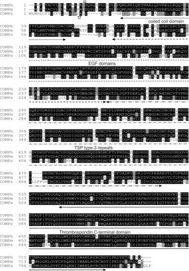
Alignment of amino acid sequences of the human, mouse and zebrafish COMP. The vertical arrows mark the potential signal peptide cleavage sites (up, zebrafish; down, human and mouse). The positions of the domains are indicated by horizontal arrows.

Phylogenetic tree of thrombospondins. Thrombospondin sequences comprising the EGF domains, TSP type-3 repeats and the C-terminal domains from zebrafish (z) and mouse (m) were aligned using the PILEUP program of the GCG package, using the default parameters. As a full-length sequence for zebrafish thrombospondin 1a was not present in the databases, we amplified the lacking N-terminal sequence by RT-PCR (see Supplementary Figure 4). The tree was constructed using the programs PROTEIN DISTANCE, Fitch-Margoliash and CONSENSE of the PHYLIP package version 3.695. Bootstrap analyses using 100 replicates were performed to show the significance. The numbers indicate the statistical weight of the individual branches. Drosophila (d) thrombospondin was used as outgroup.

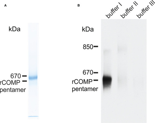
Immunoblot analysis of recombinant zebrafish Comp and Comp in extracts from zebrafish embryos. (A) Recombinant zebrafish COMP and (B) sequentially extracted proteins from whole 5 dpf zebrafish were separated in 0.5% (w/v) agarose/3% (w/v) polyacrylamide composite gels under non-reducing conditions and detected with guinea pig polyclonal antibodies specific for zebrafish Comp. Buffer I, TBS; buffer II, 50 mM Tris, pH 7.4, 10 mM EDTA, and 1M NaCl, buffer III, 50 mM Tris, pH 7.4, 10 mM EDTA and 8M urea. Recombinant rat COMP was used as a marker and the position of the pentamer is indicated.

EXPRESSION / LABELING:
Gene:
Antibody:
Fish:
Anatomical Term:
Stage: Day 5

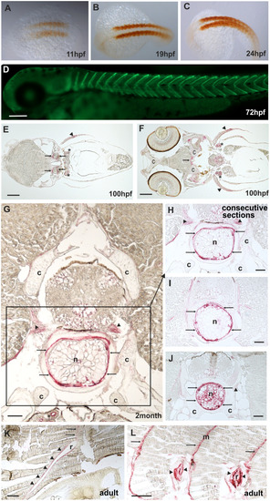
Comp distribution in zebrafish tissues at different stages of development. Immunostainings were performed using an affinity-purified rabbit antiserum directed against zebrafish Comp. (A–D), whole mount staining of 11hpf (A), 19hpf (B), 24 hpf (C) and 72 hpf (D) zebrafish. At 11-24 hpf Comp is expressed in the somites (A–C, brown peroxidase staining) and at 72 hpf in the myosepta (D, green fluorescence). E-L, immunostaining was carried out on paraffin-embedded tissue sections from 100 hpf (E, F), 2-month-old (G–J) and adult (K, L) zebrafish by alkaline phosphatase-conjugated streptavidin and Fast Red staining. In the head, at 100 hpf Comp is expressed in tendinous tissues (arrows), but not in cartilage (c) (E, F). Comp is also found in the pectoral fin (arrowheads). A transverse section through a vertebral body displays Comp in the notochord (framed) most strongly in the notochord sheath (arrows), but not in vertebral cartilage (c) of 2-month-old zebrafish (G, H, J). Consecutive transverse sections (H-J) show a variable expression around vacuolated notochord cells, most prominent at positions with a narrow notochord (J). Comp was also found in tendinous structures that attach muscles to the vertebra (G, H, J) (arrowheads). In adult zebrafish (K, L) Comp is still expressed in myosepta (m, arrows) and around ribs (r, arrowheads) where the skeletal muscles are attached. (Asterisks in E, F) Unspecific staining of kidney tubules by the secondary antibody, see also Supplementary Figure 2. Bars: 50 µm in (G–J), 100 µm in (E, F, K, L) and 200 µm in (D).

EXPRESSION / LABELING:
Gene:
Antibody:
Fish:
Anatomical Terms:
Stage Range: 1-4 somites to Adult

Mutant ΔI258D259Comp is expressed in zebrafish. (A) location of the CRISPR-Cas introduced deletion ΔI258D259 (Δ ID) in the first TSP type-3 repeat (red bar below) and comparison with mutations in chondrodysplasia patients (46) (above in red). (B) immunoblot analysis using an affinity-purified rabbit antiserum specific for zebrafish Comp (left) of direct extracts from 2-month-old wild type (wt), and heterozygous (het) or homozygous (hom) ΔI258D259Comp (Δ ID) zebrafish that were submitted to electrophoresis on 4-10% gradient SDS-polyacrylamide gels under non-reducing conditions. Ponceau staining shows equal loading (right). (C) whole mount immunofluorescence (green) staining of 72 hpf wild type (wt) (n=6), or heterozygous (het) (n=10) and homozygous (hom) (n=4) ΔI258D259Comp zebrafish mutants were performed using an affinity-purified guinea pig antiserum specific for zebrafish Comp and detected patchy Comp deposition in myosepta of heterozygous and homozygous mutants. Bar: 150 µm.

Mutant ΔI258D259Comp expression in vertebral column and muscle of zebrafish. Comp immunostainings were performed on paraffin-embedded tissue sections (A-D) from 5-month-old wild type (wt) (A, C) (n=4) and homozygous ΔI258D259Comp (B, D) (n=4) zebrafish by using affinity-purified rabbit (A-D) and guinea pig (E, F) antisera specific for zebrafish Comp, alkaline phosphatase-conjugated streptavidin and Fast Red staining (A-D) and secondary Alexa 488 conjugated antibodies (E, F). (A) In the vertebral column of 5-month-old wt zebrafish, Comp is found in the fibrocartilaginous tissue at the base of the intervertebral discs (ivd) (arrows) and lining the inner part of the bones (b) (arrowheads), (vt, vacuolated tissue). (B) In mutant ΔI258D259Comp zebrafish this expression is nearly completely lacking. However, the architecture of the vertebral column is not altered. (C) In adult wild type zebrafish Comp is still uniformly found in myosepta (m). (D) Also in adult mutant ΔI258D259Comp zebrafish Comp is found in myosepta (m) but with an irregular patchy distribution (arrowheads). (E, F) This is similar in 72 hpf zebrafish where Comp is strongly and uniformly found in myosepta (m) of wt (E) and in irregular patches (arrowheads) interrupted by a much weaker uniform staining (arrows) in ΔI258D259Comp zebrafish as shown by whole mount immunofluorescence staining using an affinity-purified guinea pig antiserum specific for zebrafish Comp (E, F) (n=100), see also Figure 6C. Bars: 100 µm in (A-D) and 10 µm in (E, F).

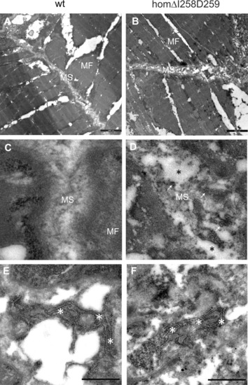
Electron microscopy of myosepta in wild type and mutant ΔI258D259Comp skeletal muscle of 72hpf zebrafish. The skeletal muscle fiber (MF) structure is comparable in wild type (wt) (A) and mutant ΔI258D259Comp (B) zebrafish while the myosepta (MS) in mutants are less dense and slightly enlarged compared to control. At higher magnification the myosepta of wild type (C) show a largely homogenous extracellular matrix (ECM) structure while myosepta of mutants (D) show disrupted (black asterisks), inhomogeneous and clustered ECM (arrows) structure with different density. The fibroblasts in wild type (E) and mutants (F) show an extensive endoplasmic reticulum (white asterisks) without obvious differences, indicating intact synthesis- active fibroblasts and no significant retention of mutant protein.

Acknowledgments
This image is the copyrighted work of the attributed author or publisher, and ZFIN has permission only to display this image to its users. Additional permissions should be obtained from the applicable author or publisher of the image. Full text @ Front Endocrinol (Lausanne)