tmem216 was widely expressed in zebrafish. In situ hybridization with tmem216 sense and antisense probes was performed on cryosections of 3- and 7-dpf zebrafish larvae. (A) Antisense probe labeling of longitudinal sections of a 3-dpf larva. tmem216 was widely expressed throughout the zebrafish, including the muscle, pronephros, brain, intestine, and liver. (B, C) Antisense probe labeling of 3-dpf zebrafish head. tmem216 was widely expressed throughout the brain and neural retina. (D) Antisense probe labeling of 7-dpf zebrafish retina. tmem216 expression persisted throughout the neural retina at 7-dpf. (E–H) Sense probe labeling of 3- and 7-dpf zebrafish cryosections. (I) RT-PCR of wildtype fish showing TMEM216 expression in newly fertilized eggs, 7-dpf larva, and 8-mpf eye, brain, and skeletal muscle. Scale bar in H: 200 µm for A, B, E, and F; 15.5 µm for C, D, G, and H.

EXPRESSION / LABELING:
Gene:
Fish:
Anatomical Terms:
Stage Range: 1-cell to Adult

Two tmem216 knockout zebrafish lines were generated by genome editing. Zebrafish knockout lines, tmem216snyΔ175 and tmem216snyR8Δ60, were generated by CRISPR/Cas9 genome editing. (A) Alignment of mutant genomic sequences with wildtype sequence from exon 3 to exon 4. The tmem216snyΔ175 line features two deletions of two stretches of DNA of 172 and three nucleotides. The tmem216snyR8Δ60 line features an eight-nucleotide repeat and deletion of two stretches of DNA of 56 and four nucleotides. Both mutations were expected to result in frameshift. (B) Protein sequence alignment between wildtype TMEM216, and the expected TMEM216SNYΔ175 and TMEM216SNYR8Δ60 mutant proteins. Note both mutations result in the loss of all four transmembrane domains. (C) RT-PCR of wild-type, heterozygous, and homozygous zebrafish, showing that homozygous knockouts express only mutant mRNA.

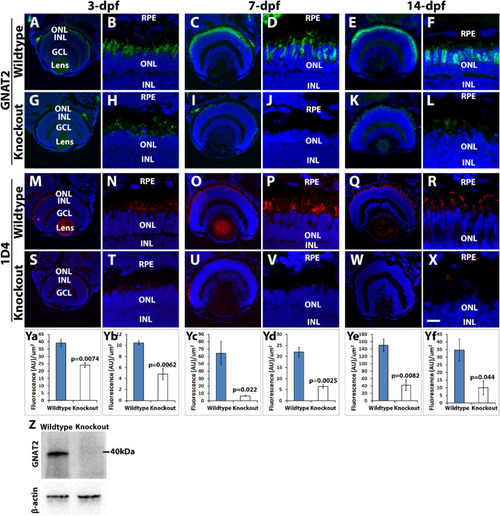
Cone outer segment generation is reduced in tmem216 knockout fish. Cryosections of whole zebrafish heads were stained with cone OS markers GNAT2 (green) and 1D4 (red) at 3-, 7- and 14-dpf. All sections were counterstained with DAPI to visualize nuclei. Knockout panels shown were from the tmem216snyR8Δ60 line. Similar results were observed in the tmem216snyΔ175 line. (A–F) GNAT2 immunostaining of wild-type retina at 3-, 7-, and 14-dpf, respectively. (G–L) GNAT2 staining of tmem216snyR8Δ60 homozygous retina at 3-, 7-, and 14-dpf, respectively. (M–R) 1D4 immunostaining of wild-type retina at 3-, 7-, and 14-dpf, respectively. (S–X) 1D4 labeling of tmem216snyR8Δ60 homozygous retina at 3-, 7-, and 14-dpf, respectively. (Ya, Yc, Ye) Quantification of GNAT2 fluorescence intensity between wildtype and knockout retinas at 3-, 7-, and 14-dpf, respectively. (Yb, Yd, Yf) Quantification of 1D4 fluorescence intensity between wildtype and tmem216snyR8Δ60 homozygous retina at 3-, 7- and 14-dpf, respectively. Note that GNAT2 and 1D4 immunoreactivity was significantly reduced at 3-, 7- and 14-dpf (n = 3, Student's t-test). (Z) Western blotting using anti-GNAT2 was performed on whole head lysates collected from three 7-dpf wildtype and tmem216snyR8Δ60 homozygous zebrafish larvae. The intensity ratio of GNAT2/-β-actin between wildtype and knockout was reduced from 0.88 to 0.27. Scale bar in X: 55 µm for A, G, M, and S; 110 µm for C, I, E, K, O, U, Q, and W; 6 µm for B, H, D, J, F, L, N, T, P, V, R, and X. GCL, ganglion cell layer; INL, inner nuclear layer; ONL, outer nuclear layer; RPE, retinal pigment epithelium.

Mislocalization of cone outer segment proteins in tmem216 knockout photoreceptors. Cryosections were immunostained with GNAT2 (green) and 1D4 (red). (A, C) GNAT2 staining for wildtype and tmem216snyR8Δ60 homozygous retina. Similar phenotypes were observed in the tmem216snyΔ175 homozygous retina. GNAT2 was normally found in the cone outer segments (arrow in A) in the wildtype retina. Mislocalization of GNAT2 reactivity to the cone cell body was frequently found in the knockout fish (arrowheads in C). (B, D) 1D4 staining of wildtype and knockout retina. 1D4 reactivity was labeling long double cone outer segments in the wildtype (arrow in B). 1D4 reactivity was frequently found around the cell bodies of the knockout retina (arrowhead, in D). Scale bar in D: 6 µm.

Rod outer segments were reduced in tmem216 knockout zebrafish. Retinal sections were stained with rhodopsin marker, 4D2 (red), and counterstained with DAPI to label nuclei. Knockout images shown represent the tmem216snyΔ175 retina, similar phenotypes were observed in the tmem216snyR8Δ60 homozygous retina. (A–F) Wildtype at 3-, 7-, and 14-dpf, respectively. (G–L) tmem216snyΔ175 homozygous at 3-, 7-, and 14-dpf, respectively. (M–O) 4D2 immunofluorescence intensity was significantly reduced at 3-, 7-, and 14-dpf in tmem216snyΔ175 homozygous retina (n = 3, Student's t-test). Scale bar in L: 55 µm for A and G; 110 µm for C, I, E, and K; 6 µm for B, H, D, J, F, and L.

Defective organization of F-actin in tmem216 knockout photoreceptors. Cryosections were immunostained with 4D2 (red). Some sections were stained with Phalloidin-RITC for F-actin (red). (A, C) 4D2 staining of wildtype and tmem216snyΔ175 homozygous retina. 4D2 labels rod outer segments in the wildtype zebrafish (arrow in A); however, its reactivity was frequently mislocalized to the rod cell body in the knockouts (arrowheads in C). (B, D) Phalloidin staining in wildtype and tmem216snyΔ175 homozygous retina. Vertically oriented F-actin in the wildtype (arrows in B) were disrupted in tmem216snyΔ175 homozygous retina (arrowheads in D). Scale bar in H: 6 µm.

TUNEL-positive photoreceptor nuclei were increased in tmem216 knockouts. Cryosections from 7-dpf zebrafish were stained by TUNEL assay (green) and anti-CHOP (Proteintech), counter stained with DAPI. (A) Wildtype. (B, C) tmem216snyR8Δ60 homozygous retina. Note TUNEL-positive DAPI-labeled rod (arrow in B) and cone (arrow in C) nuclei. (D) Quantification of TUNEL-positive nuclei. Number of TUNEL-positive nuclei were significantly increased in the outer nuclear layer of tmem216snyR8Δ60 homozygous animals (n = 3, Student's t-test); similar phenotype was observed in tmem216snyΔ175 animals. (E, F) CHOP staining of wildtype and knockout retina. Immunoreactivity of CHOP antibody was similar between wildtype and tmem216snyR8Δ60 homozygous retina. Scale bar in A: 6 µm for A, E, and F; 8 µm for B and C.

Length of photoreceptor axonemes was reduced in knockout zebrafish. Cryosections were immunostained with axoneme marker, acetylated α-tubulin (red), and connecting ciliary region markers CC2D2A, TMEM231, and EYS (green). Sections were then counterstained with DAPI to visualize nuclei. (A, B) Acetylated α-tubulin (red, arrow) and CC2D2A (green, arrowhead) double immuno-labeling of 7dpf wildtype and tmem216snyΔ175 homozygous retina. (C, D) Acetylated α-tubulin (red, arrow) and TMEM231 (green, arrowhead) double immuno-labeling of 7dpf wildtype and tmem216snyΔ175 homozygous retina. (E, F) Acetylated α-tubulin (red, arrow) and EYS (green, arrowhead) double immuno-labeling of 7dpf wildtype and tmem216snyΔ175 homozygous retina. Note the reduction in axoneme length in the knockout zebrafish (F, inset). (J, K) Acetylated α-tubulin and EYS double immuno-labeling of 14-dpf wildtype and tmem216snyΔ175 homozygous retina. (G–I) Quantification of 7-dpf axoneme number and length. Note the reduction in average axoneme length (n = 3, Student's t-test) and the shift of axoneme lengths toward lower length bins in the knockout. (L–N) Quantification of 14-dpf axoneme number and length (n = 3, Student's t-test). Scale bar in K: 6 µm for A–F, J, and K; 1.5 µm for insets.

tmem216 knockouts exhibit abnormal outer segment disc morphology. Eyes from 7-dpf wildtype and tmem216snyR8Δ60 zebrafish were processed for transmission EM. (A–D) Wildtype retina. Note that the outer segments with uniform discs. (E–I) tmem216snyR8Δ60 homozygous retina. Although photoreceptors in the tmem216snyR8Δ60 homozygous retina elaborated cilia (asterisk in F), the outer segments of these photoreceptors exhibit multiple defects. Abnormalities manifested as large vacuoles within the outer segment (asterisks in G and H), large vacuoles at the base of the outer segment (asterisks in I), shortened discs (arrows in G), abnormal disc morphology (arrowhead in G). Scale bar in I: 4 µm for A and E; 400 nm for B, D, and F; 200 nm for C, G, H, and I. BB, basal body; CP, ciliary pocket; IS, inner segment; OS, outer segment.

ZFIN is incorporating published figure images and captions as part of an ongoing project. Figures from some publications have not yet been curated, or are not available for display because of copyright restrictions.

Acknowledgments
This image is the copyrighted work of the attributed author or publisher, and ZFIN has permission only to display this image to its users. Additional permissions should be obtained from the applicable author or publisher of the image. Full text @ Invest. Ophthalmol. Vis. Sci.